3HSY

High resolution structure of a dimeric GluR2 N-terminal domain (NTD)


Experimental Data Snapshot

  • Method: X-RAY DIFFRACTION
  • Resolution: 1.75 Å
  • R-Value Free: 0.205 
  • R-Value Work: 0.178 
  • R-Value Observed: 0.178 

wwPDB Validation   3D Report Full Report


Ligand Structure Quality Assessment 


This is version 2.0 of the entry. See complete history


Literature

Subunit-selective N-terminal domain associations organize the formation of AMPA receptor heteromers

Rossmann, M.Sukumaran, M.Penn, A.C.Veprintsev, D.B.Babu, M.M.Greger, I.H.

(2011) EMBO J 30: 959-971

  • DOI: https://doi.org/10.1038/emboj.2011.16
  • Primary Citation of Related Structures:  
    3HSY, 3N6V

  • PubMed Abstract: 

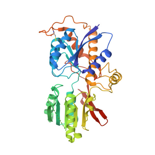
    The assembly of AMPA-type glutamate receptors (AMPARs) into distinct ion channel tetramers ultimately governs the nature of information transfer at excitatory synapses. How cells regulate the formation of diverse homo- and heteromeric AMPARs is unknown. Using a sensitive biophysical approach, we show that the extracellular, membrane-distal AMPAR N-terminal domains (NTDs) orchestrate selective routes of heteromeric assembly via a surprisingly wide spectrum of subunit-specific association affinities. Heteromerization is dominant, occurs at the level of the dimer, and results in a preferential incorporation of the functionally critical GluA2 subunit. Using a combination of structure-guided mutagenesis and electrophysiology, we further map evolutionarily variable hotspots in the NTD dimer interface, which modulate heteromerization capacity. This 'flexibility' of the NTD not only explains why heteromers predominate but also how GluA2-lacking, Ca(2+)-permeable homomers could form, which are induced under specific physiological and pathological conditions. Our findings reveal that distinct NTD properties set the stage for the biogenesis of functionally diverse pools of homo- and heteromeric AMPAR tetramers.


  • Organizational Affiliation

    Neurobiology Division, MRC Laboratory of Molecular Biology, Cambridge, UK.


Macromolecules
Find similar proteins by:  (by identity cutoff)  |  3D Structure
Entity ID: 1
MoleculeChains Sequence LengthOrganismDetailsImage
Glutamate receptor 2
A, B
376Rattus norvegicusMutation(s): 0 
Gene Names: Gria2Glur2
UniProt
Find proteins for P19491 (Rattus norvegicus)
Explore P19491 
Go to UniProtKB:  P19491
Entity Groups  
Sequence Clusters30% Identity50% Identity70% Identity90% Identity95% Identity100% Identity
UniProt GroupP19491
Sequence Annotations
Expand
  • Reference Sequence
Oligosaccharides

Help

Entity ID: 2
MoleculeChains Length2D Diagram Glycosylation3D Interactions
beta-D-mannopyranose-(1-4)-2-acetamido-2-deoxy-beta-D-glucopyranose-(1-4)-2-acetamido-2-deoxy-beta-D-glucopyranose
C
3N-Glycosylation
Glycosylation Resources
GlyTouCan:  G15407YE
GlyCosmos:  G15407YE
GlyGen:  G15407YE
Experimental Data & Validation

Experimental Data

  • Method: X-RAY DIFFRACTION
  • Resolution: 1.75 Å
  • R-Value Free: 0.205 
  • R-Value Work: 0.178 
  • R-Value Observed: 0.178 
  • Space Group: P 21 21 21
Unit Cell:
Length ( Å )Angle ( ˚ )
a = 82.452α = 90
b = 93.695β = 90
c = 101.466γ = 90
Software Package:
Software NamePurpose
SCALAdata processing
SHARPphasing
PHENIXrefinement
PDB_EXTRACTdata extraction
ADSCdata collection
XDSdata reduction
SCALAdata scaling
autoSHARPphasing

Structure Validation

View Full Validation Report


Ligand Structure Quality Assessment 


Entry History 

Deposition Data

Revision History  (Full details and data files)

  • Version 1.0: 2010-06-16
    Type: Initial release
  • Version 1.1: 2011-07-13
    Changes: Version format compliance
  • Version 2.0: 2020-07-29
    Type: Remediation
    Reason: Carbohydrate remediation
    Changes: Atomic model, Data collection, Derived calculations, Structure summary