3HRW

Crystal structure of hemoglobin from mouse (Mus musculus)at 2.8


Experimental Data Snapshot

  • Method: X-RAY DIFFRACTION
  • Resolution: 2.80 Å
  • R-Value Free: 0.306 
  • R-Value Work: 0.243 
  • R-Value Observed: 0.245 

wwPDB Validation   3D Report Full Report


Ligand Structure Quality Assessment 


This is version 1.3 of the entry. See complete history


Literature

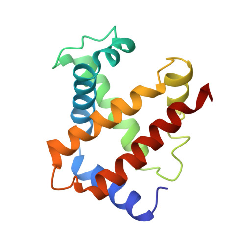
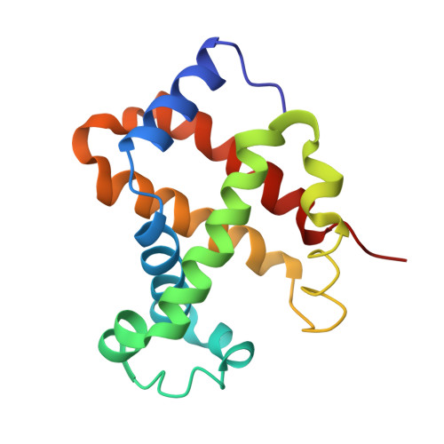
Crystal structure of hemoglobin from mouse (Mus musculus) compared with those from other small animals and humans.

Sundaresan, S.S.Ramesh, P.Shobana, N.Vinuchakkaravarthy, T.Yasien, S.Ponnuswamy, M.N.G.

(2021) Acta Crystallogr F Struct Biol Commun 77: 113-120

  • DOI: https://doi.org/10.1107/S2053230X2100306X
  • Primary Citation of Related Structures:  
    3HRW

  • PubMed Abstract: 

    Mice (Mus musculus) are nocturnal small animals belonging to the rodent family that live in burrows, an environment in which significantly high CO 2 levels prevail. It is expected that mouse hemoglobin (Hb) plays an important role in their adaptation to living in such a high-CO 2 environment, while many other species cannot. In the present study, mouse Hb was purified and crystallized at a physiological pH of 7 in the orthorhombic space group P2 1 2 1 2 1 ; the crystals diffracted to 2.8 Å resolution. The primary amino-acid sequence and crystal structure of mouse Hb were compared with those of mammalian Hbs in order to investigate the structure-function relationship of mouse Hb. Differences were observed from guinea pig Hb in terms of amino-acid sequence and from cat Hb in overall structure (in terms of r.m.s.d.). The difference in r.m.s.d. from cat Hb may be due to the existence of the molecule in a conformation other than the R-state. Analysis of tertiary- and quaternary-structural features, the α1β2 interface region and the heme environment without any ligands in all four heme groups showed that mouse methemoglobin is in an intermediate state between the R-state and the T-state that is much closer to the R-state conformation.


  • Organizational Affiliation

    Department of Physics, Sir Theagaraya College, Chennai 600 021, India.


Macromolecules
Find similar proteins by:  (by identity cutoff)  |  3D Structure
Entity ID: 1
MoleculeChains Sequence LengthOrganismDetailsImage
Hemoglobin subunit alpha
A, C
141Mus musculusMutation(s): 0 
UniProt
Find proteins for P01942 (Mus musculus)
Explore P01942 
Go to UniProtKB:  P01942
Entity Groups  
Sequence Clusters30% Identity50% Identity70% Identity90% Identity95% Identity100% Identity
UniProt GroupP01942
Sequence Annotations
Expand
  • Reference Sequence
Find similar proteins by:  (by identity cutoff)  |  3D Structure
Entity ID: 2
MoleculeChains Sequence LengthOrganismDetailsImage
Hemoglobin subunit beta-1
B, D
146Mus musculusMutation(s): 0 
UniProt
Find proteins for P02088 (Mus musculus)
Explore P02088 
Go to UniProtKB:  P02088
Entity Groups  
Sequence Clusters30% Identity50% Identity70% Identity90% Identity95% Identity100% Identity
UniProt GroupP02088
Sequence Annotations
Expand
  • Reference Sequence
Experimental Data & Validation

Experimental Data

  • Method: X-RAY DIFFRACTION
  • Resolution: 2.80 Å
  • R-Value Free: 0.306 
  • R-Value Work: 0.243 
  • R-Value Observed: 0.245 
  • Space Group: P 21 21 21
Unit Cell:
Length ( Å )Angle ( ˚ )
a = 53.081α = 90
b = 65.94β = 90
c = 150.931γ = 90
Software Package:
Software NamePurpose
MAR345dtbdata collection
PHASESphasing
REFMACrefinement
AUTOMARdata reduction
SCALEPACKdata scaling

Structure Validation

View Full Validation Report


Ligand Structure Quality Assessment 


Entry History 

Deposition Data

Revision History  (Full details and data files)

  • Version 1.0: 2009-06-23
    Type: Initial release
  • Version 1.1: 2011-07-13
    Changes: Version format compliance
  • Version 1.2: 2021-04-21
    Changes: Database references, Derived calculations
  • Version 1.3: 2023-11-01
    Changes: Data collection, Database references, Refinement description