3HQG

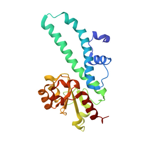
Crystal structure of restriction endonuclease EcoRII catalytic C-terminal domain in complex with cognate DNA


Experimental Data Snapshot

  • Method: X-RAY DIFFRACTION
  • Resolution: 2.60 Å
  • R-Value Free: 0.293 
  • R-Value Work: 0.236 
  • R-Value Observed: 0.242 

wwPDB Validation   3D Report Full Report


This is version 1.2 of the entry. See complete history


Literature

Structural mechanisms for the 5'-CCWGG sequence recognition by the N- and C-terminal domains of EcoRII.

Golovenko, D.Manakova, E.Tamulaitiene, G.Grazulis, S.Siksnys, V.

(2009) Nucleic Acids Res 37: 6613-6624

  • DOI: https://doi.org/10.1093/nar/gkp699
  • Primary Citation of Related Structures:  
    3HQF, 3HQG

  • PubMed Abstract: 

    EcoRII restriction endonuclease is specific for the 5'-CCWGG sequence (W stands for A or T); however, it shows no activity on a single recognition site. To activate cleavage it requires binding of an additional target site as an allosteric effector. EcoRII dimer consists of three structural units: a central catalytic core, made from two copies of the C-terminal domain (EcoRII-C), and two N-terminal effector DNA binding domains (EcoRII-N). Here, we report DNA-bound EcoRII-N and EcoRII-C structures, which show that EcoRII combines two radically different structural mechanisms to interact with the effector and substrate DNA. The catalytic EcoRII-C dimer flips out the central T:A base pair and makes symmetric interactions with the CC:GG half-sites. The EcoRII-N effector domain monomer binds to the target site asymmetrically in a single defined orientation which is determined by specific hydrogen bonding and van der Waals interactions with the central T:A pair in the major groove. The EcoRII-N mode of the target site recognition is shared by the large class of higher plant transcription factors of the B3 superfamily.


  • Organizational Affiliation

    Institute of Biotechnology, Graiciuno 8, LT-02241 Vilnius, Lithuania.


Macromolecules

Find similar proteins by:  (by identity cutoff)  |  3D Structure
Entity ID: 1
MoleculeChains Sequence LengthOrganismDetailsImage
Type-2 restriction enzyme EcoRII222Escherichia coliMutation(s): 0 
Gene Names: ECORIIecoRIIR
EC: 3.1.21.4
UniProt
Find proteins for P14633 (Escherichia coli)
Explore P14633 
Go to UniProtKB:  P14633
Entity Groups  
Sequence Clusters30% Identity50% Identity70% Identity90% Identity95% Identity100% Identity
UniProt GroupP14633
Sequence Annotations
Expand
  • Reference Sequence

Find similar nucleic acids by:  Sequence   |   3D Structure  

Entity ID: 2
MoleculeChains LengthOrganismImage
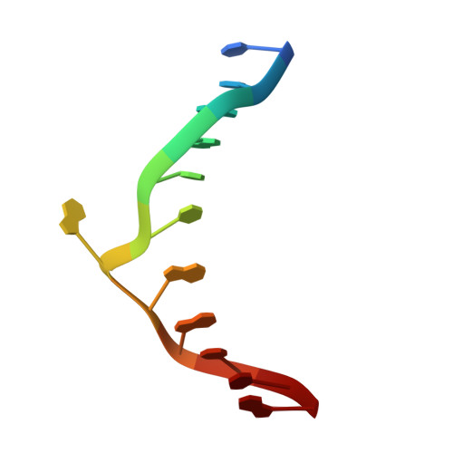
5'-D(*TP*CP*GP*AP*CP*CP*AP*GP*GP*CP*TP*A)-3'12N/A
Sequence Annotations
Expand
  • Reference Sequence

Find similar nucleic acids by:  Sequence   |   3D Structure  

Entity ID: 3
MoleculeChains LengthOrganismImage
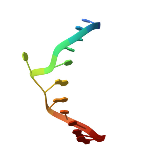
5'-D(*TP*AP*GP*CP*CP*TP*GP*GP*TP*CP*GP*A)-3'12N/A
Sequence Annotations
Expand
  • Reference Sequence
Small Molecules
Ligands 1 Unique
IDChains Name / Formula / InChI Key2D Diagram3D Interactions
GOL
Query on GOL

Download Ideal Coordinates CCD File 
D [auth A]GLYCEROL
C3 H8 O3
PEDCQBHIVMGVHV-UHFFFAOYSA-N
Experimental Data & Validation

Experimental Data

  • Method: X-RAY DIFFRACTION
  • Resolution: 2.60 Å
  • R-Value Free: 0.293 
  • R-Value Work: 0.236 
  • R-Value Observed: 0.242 
  • Space Group: P 21 21 2
Unit Cell:
Length ( Å )Angle ( ˚ )
a = 77.054α = 90
b = 57.971β = 90
c = 61.01γ = 90
Software Package:
Software NamePurpose
REFMACrefinement
AMoREphasing
MOSFLMdata reduction
SCALAdata scaling
PDB_EXTRACTdata extraction

Structure Validation

View Full Validation Report


Entry History 

Deposition Data

Revision History  (Full details and data files)

  • Version 1.0: 2009-09-22
    Type: Initial release
  • Version 1.1: 2011-07-13
    Changes: Advisory, Refinement description, Version format compliance
  • Version 1.2: 2023-09-06
    Changes: Data collection, Database references, Derived calculations, Refinement description