


3H5K
Crystal structure of the ribosome inactivating protein PDL1
- PDB DOI: https://doi.org/10.2210/pdb3H5K/pdb
- Classification: HYDROLASE
- Organism(s): Phytolacca dioica
- Mutation(s): No
- Deposited: 2009-04-22 Released: 2009-10-13
Experimental Data Snapshot
- Method: X-RAY DIFFRACTION
- Resolution: 1.45 Å
- R-Value Free: 0.242
- R-Value Work: 0.196
- R-Value Observed: 0.198

This is version 1.2 of the entry. See complete history.

Macromolecules
Find similar proteins by:
(by identity cutoff) | 3D Structure
Entity ID: 1 | |||||
|---|---|---|---|---|---|
| Molecule | Chains | Sequence Length | Organism | Details | Image |
| Ribosome-inactivating protein PD-L1/PD-L2 | 261 | Phytolacca dioica | Mutation(s): 0 EC: 3.2.2.22 | 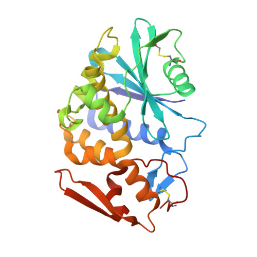 | |
UniProt | |||||
Find proteins for P84853 (Phytolacca dioica) Explore P84853 Go to UniProtKB: P84853 | |||||
Entity Groups | |||||
| Sequence Clusters | 30% Identity50% Identity70% Identity90% Identity95% Identity100% Identity | ||||
| UniProt Group | P84853 | ||||
Sequence AnnotationsExpand | |||||
| |||||
Small Molecules
| Ligands 2 Unique | |||||
|---|---|---|---|---|---|
| ID | Chains | Name / Formula / InChI Key | 2D Diagram | 3D Interactions | |
| NAG Query on NAG | C [auth A], E [auth B] | 2-acetamido-2-deoxy-beta-D-glucopyranose C8 H15 N O6 OVRNDRQMDRJTHS-FMDGEEDCSA-N |  | ||
| EDO Query on EDO | D [auth A], F [auth B] | 1,2-ETHANEDIOL C2 H6 O2 LYCAIKOWRPUZTN-UHFFFAOYSA-N |  | ||
Experimental Data & Validation
Experimental Data
- Method: X-RAY DIFFRACTION
- Resolution: 1.45 Å
- R-Value Free: 0.242
- R-Value Work: 0.196
- R-Value Observed: 0.198
- Space Group: C 1 2 1
Unit Cell:
| Length ( Å ) | Angle ( ˚ ) |
|---|---|
| a = 161.011 | α = 90 |
| b = 34.735 | β = 127.99 |
| c = 120.63 | γ = 90 |
| Software Name | Purpose |
|---|---|
| HKL-2000 | data collection |
| REMO | model building |
| REFMAC | refinement |
| HKL-2000 | data reduction |
| HKL-2000 | data scaling |
| REMO | phasing |
Entry History
Deposition Data
- Released Date: 2009-10-13 Deposition Author(s): Ruggiero, A., Di Maro, A., Severino, V., Chambery, A., Berisio, R.
Revision History (Full details and data files)
- Version 1.0: 2009-10-13
Type: Initial release - Version 1.1: 2011-07-13
Changes: Non-polymer description, Version format compliance - Version 1.2: 2020-07-29
Type: Remediation
Reason: Carbohydrate remediation
Changes: Data collection, Derived calculations, Structure summary
