


Crystal Structures of Two P60-Family Antigens from Mycobacterium Avium Paratuberculosis
Ramyar, K.X., Lingle, C.K., McWhorter, W.J., Bouyain, S., Bannantine, J.P., Geisbrecht, B.V.To be published.
Experimental Data Snapshot
wwPDB Validation 3D Report Full Report

Entity ID: 1 | |||||
|---|---|---|---|---|---|
| Molecule | Chains | Sequence Length | Organism | Details | Image |
| Putative uncharacterized protein | 142 | Mycobacterium avium subsp. paratuberculosis | Mutation(s): 0 Gene Names: MAP1272c, MAP_1272c | 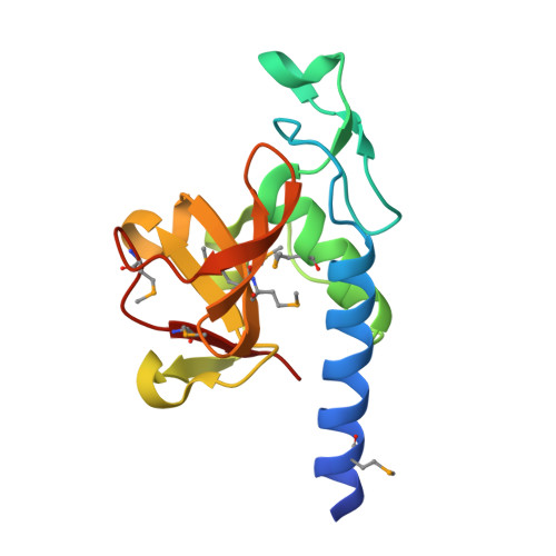 | |
UniProt | |||||
Find proteins for Q740S0 (Mycolicibacterium paratuberculosis (strain ATCC BAA-968 / K-10)) Explore Q740S0 Go to UniProtKB: Q740S0 | |||||
Entity Groups | |||||
| Sequence Clusters | 30% Identity50% Identity70% Identity90% Identity95% Identity100% Identity | ||||
| UniProt Group | Q740S0 | ||||
Sequence AnnotationsExpand | |||||
| |||||
| Ligands 2 Unique | |||||
|---|---|---|---|---|---|
| ID | Chains | Name / Formula / InChI Key | 2D Diagram | 3D Interactions | |
| SO4 Query on SO4 | B [auth A], C [auth A] | SULFATE ION O4 S QAOWNCQODCNURD-UHFFFAOYSA-L |  | ||
| EDO Query on EDO | D [auth A], E [auth A] | 1,2-ETHANEDIOL C2 H6 O2 LYCAIKOWRPUZTN-UHFFFAOYSA-N |  | ||
| Modified Residues 1 Unique | |||||
|---|---|---|---|---|---|
| ID | Chains | Type | Formula | 2D Diagram | Parent |
| MSE Query on MSE | A | L-PEPTIDE LINKING | C5 H11 N O2 Se |  | MET |
| Length ( Å ) | Angle ( ˚ ) |
|---|---|
| a = 34.715 | α = 90 |
| b = 53.212 | β = 102.95 |
| c = 38.325 | γ = 90 |
| Software Name | Purpose |
|---|---|
| DENZO | data reduction |
| SCALEPACK | data scaling |
| PHASES | phasing |
| RESOLVE | phasing |
| PHENIX | refinement |
| PDB_EXTRACT | data extraction |
| HKL-2000 | data collection |
| PHENIX | phasing |
RCSB PDB (citation) is hosted by
RCSB PDB is a member of the

