3GGC

Human hypoxanthine-guanine phosphoribosyltransferase in complex with 9-(2-phosphonoethoxyethyl)hypoxanthine


Experimental Data Snapshot

  • Method: X-RAY DIFFRACTION
  • Resolution: 2.78 Å
  • R-Value Free: 0.285 
  • R-Value Work: 0.228 
  • R-Value Observed: 0.231 

wwPDB Validation   3D Report Full Report


Ligand Structure Quality Assessment 


This is version 1.2 of the entry. See complete history


Literature

Inhibition of hypoxanthine-guanine phosphoribosyltransferase by acyclic nucleoside phosphonates: a new class of antimalarial therapeutics.

Keough, D.T.Hockova, D.Holy, A.Naesens, L.M.Skinner-Adams, T.S.Jersey, J.Guddat, L.W.

(2009) J Med Chem 52: 4391-4399

  • DOI: https://doi.org/10.1021/jm900267n
  • Primary Citation of Related Structures:  
    3GEP, 3GGC, 3GGJ

  • PubMed Abstract: 

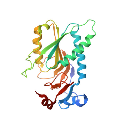
    The purine salvage enzyme hypoxanthine-guanine-xanthine phosphoribosyltransferase (HGXPRT) is essential for purine nucleotide and hence nucleic acid synthesis in the malaria parasite, Plasmodium falciparum. Acyclic nucleoside phosphonates (ANPs) are analogues of the nucleotide product of the reaction, comprising a purine base joined by a linker to a phosphonate moiety. K(i) values for 19 ANPs were determined for Pf HGXPRT and the corresponding human enzyme, HGPRT. Values for Pf HGXPRT were as low as 100 nM, with selectivity for the parasite enzyme of up to 58. Structures of human HGPRT in complex with three ANPs are reported. On binding, a large mobile loop in the free enzyme moves to partly cover the active site. For three ANPs, the IC(50) values for Pf grown in cell culture were 1, 14, and 46 microM, while the cytotoxic concentration for the first compound was 489 microM. These results provide a basis for the design of potent and selective ANP inhibitors of Pf HGXPRT as antimalarial drug leads.


  • Organizational Affiliation

    The School of Chemistry and Molecular Biosciences, The University of Queensland, Brisbane, 4072 QLD, Australia.


Macromolecules
Find similar proteins by:  (by identity cutoff)  |  3D Structure
Entity ID: 1
MoleculeChains Sequence LengthOrganismDetailsImage
Hypoxanthine-guanine phosphoribosyltransferase
A, B
217Homo sapiensMutation(s): 0 
Gene Names: HPRTHPRT1HPT
EC: 2.4.2.8
UniProt & NIH Common Fund Data Resources
Find proteins for P00492 (Homo sapiens)
Explore P00492 
Go to UniProtKB:  P00492
PHAROS:  P00492
GTEx:  ENSG00000165704 
Entity Groups  
Sequence Clusters30% Identity50% Identity70% Identity90% Identity95% Identity100% Identity
UniProt GroupP00492
Sequence Annotations
Expand
  • Reference Sequence
Small Molecules
Ligands 1 Unique
IDChains Name / Formula / InChI Key2D Diagram3D Interactions
H26
Query on H26

Download Ideal Coordinates CCD File 
C [auth A],
D [auth B]
{2-[2-(6-oxo-1,6-dihydro-9H-purin-9-yl)ethoxy]ethyl}phosphonic acid
C9 H13 N4 O5 P
ARUATFPHVPUVFJ-UHFFFAOYSA-N
Binding Affinity Annotations 
IDSourceBinding Affinity
H26 PDBBind:  3GGC Ki: 3600 (nM) from 1 assay(s)
Experimental Data & Validation

Experimental Data

  • Method: X-RAY DIFFRACTION
  • Resolution: 2.78 Å
  • R-Value Free: 0.285 
  • R-Value Work: 0.228 
  • R-Value Observed: 0.231 
  • Space Group: P 21 21 2
Unit Cell:
Length ( Å )Angle ( ˚ )
a = 111.284α = 90
b = 72.775β = 90
c = 50.993γ = 90
Software Package:
Software NamePurpose
CrystalCleardata collection
REFMACrefinement
CrystalCleardata reduction
CrystalCleardata scaling

Structure Validation

View Full Validation Report


Ligand Structure Quality Assessment 


Entry History 

Deposition Data

Revision History  (Full details and data files)

  • Version 1.0: 2009-08-18
    Type: Initial release
  • Version 1.1: 2011-07-13
    Changes: Advisory, Version format compliance
  • Version 1.2: 2023-11-01
    Changes: Data collection, Database references, Derived calculations, Refinement description