3GD3

Crystal structure of a naturally folded murine apoptosis inducing factor


Experimental Data Snapshot

  • Method: X-RAY DIFFRACTION
  • Resolution: 2.95 Å
  • R-Value Free: 0.300 
  • R-Value Work: 0.249 
  • R-Value Observed: 0.251 

wwPDB Validation   3D Report Full Report


Ligand Structure Quality Assessment 


This is version 1.2 of the entry. See complete history


Literature

Redox-linked conformational dynamics in apoptosis-inducing factor

Sevrioukova, I.F.

(2009) J Mol Biol 390: 924-938

  • DOI: https://doi.org/10.1016/j.jmb.2009.05.013
  • Primary Citation of Related Structures:  
    3GD3, 3GD4

  • PubMed Abstract: 

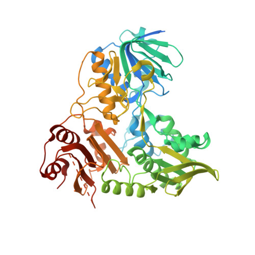
    Apoptosis-inducing factor (AIF) is a bifunctional mitochondrial flavoprotein critical for energy metabolism and induction of caspase-independent apoptosis, whose exact role in normal mitochondria remains unknown. Upon reduction with NADH, AIF undergoes dimerization and forms tight, long-lived FADH(2)-NAD charge-transfer complexes (CTC) that are proposed to be functionally important. To obtain a deeper insight into structure/function relations and redox mechanism of this vitally important protein, we determined the X-ray structures of oxidized and NADH-reduced forms of naturally folded recombinant murine AIF. Our structures reveal that CTC with the pyridine nucleotide is stabilized by (i) pi-stacking interactions between coplanar nicotinamide, isoalloxazine, and Phe309 rings; (ii) rearrangement of multiple aromatic residues in the C-terminal domain, likely serving as an electron delocalization site; and (iii) an extensive hydrogen-bonding network involving His453, a key residue that undergoes a conformational switch to directly interact with and optimally orient the nicotinamide for charge transfer. Via the His453-containing peptide, redox changes in the active site are transmitted to the surface, promoting AIF dimerization and restricting access to a primary nuclear localization signal through which the apoptogenic form is transported to the nucleus. Structural findings agree with biochemical data and support the hypothesis that both normal and apoptogenic functions of AIF are controlled by NADH.


  • Organizational Affiliation

    Department of Molecular Biology and Biochemistry, University of California, Irvine, 92697-3900, USA. sevrioui@uci.edu


Macromolecules
Find similar proteins by:  (by identity cutoff)  |  3D Structure
Entity ID: 1
MoleculeChains Sequence LengthOrganismDetailsImage
Apoptosis-inducing factor 1, mitochondrial
A, B, C, D
535Mus musculusMutation(s): 0 
Gene Names: AifAifm1Pdcd8
UniProt
Find proteins for Q9Z0X1 (Mus musculus)
Explore Q9Z0X1 
Go to UniProtKB:  Q9Z0X1
Entity Groups  
Sequence Clusters30% Identity50% Identity70% Identity90% Identity95% Identity100% Identity
UniProt GroupQ9Z0X1
Sequence Annotations
Expand
  • Reference Sequence

Find similar proteins by:  Sequence   |   3D Structure  

Entity ID: 2
MoleculeChains Sequence LengthOrganismDetailsImage
Apoptosis-inducing factor 1, mitochondrial
E, F
9Mus musculusMutation(s): 0 
Gene Names: AifAifm1Pdcd8
Sequence Annotations
Expand
  • Reference Sequence
Experimental Data & Validation

Experimental Data

  • Method: X-RAY DIFFRACTION
  • Resolution: 2.95 Å
  • R-Value Free: 0.300 
  • R-Value Work: 0.249 
  • R-Value Observed: 0.251 
  • Space Group: P 21 21 21
Unit Cell:
Length ( Å )Angle ( ˚ )
a = 75.05α = 90
b = 80.29β = 90
c = 419.64γ = 90
Software Package:
Software NamePurpose
HKL-2000data collection
PHASERphasing
REFMACrefinement
d*TREKdata reduction
d*TREKdata scaling

Structure Validation

View Full Validation Report


Ligand Structure Quality Assessment 


Entry History 

Deposition Data

Revision History  (Full details and data files)

  • Version 1.0: 2009-05-19
    Type: Initial release
  • Version 1.1: 2011-07-13
    Changes: Version format compliance
  • Version 1.2: 2023-09-06
    Changes: Data collection, Database references, Derived calculations, Refinement description