3GA8

Structure of the N-terminal domain of the E. coli protein MqsA (YgiT/b3021)


Experimental Data Snapshot

  • Method: X-RAY DIFFRACTION
  • Resolution: 1.70 Å
  • R-Value Free: 0.186 
  • R-Value Work: 0.165 
  • R-Value Observed: 0.166 

wwPDB Validation   3D Report Full Report


This is version 1.2 of the entry. See complete history


Literature

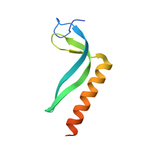
Three dimensional structure of the MqsR:MqsA complex: a novel TA pair comprised of a toxin homologous to RelE and an antitoxin with unique properties.

Brown, B.L.Grigoriu, S.Kim, Y.Arruda, J.M.Davenport, A.Wood, T.K.Peti, W.Page, R.

(2009) PLoS Pathog 5: e1000706-e1000706

  • DOI: https://doi.org/10.1371/journal.ppat.1000706
  • Primary Citation of Related Structures:  
    3FMY, 3GA8, 3GN5, 3HI2

  • PubMed Abstract: 

    One mechanism by which bacteria survive environmental stress is through the formation of bacterial persisters, a sub-population of genetically identical quiescent cells that exhibit multidrug tolerance and are highly enriched in bacterial toxins. Recently, the Escherichia coli gene mqsR (b3022) was identified as the gene most highly upregulated in persisters. Here, we report multiple individual and complex three-dimensional structures of MqsR and its antitoxin MqsA (B3021), which reveal that MqsR:MqsA form a novel toxin:antitoxin (TA) pair. MqsR adopts an alpha/beta fold that is homologous with the RelE/YoeB family of bacterial ribonuclease toxins. MqsA is an elongated dimer that neutralizes MqsR toxicity. As expected for a TA pair, MqsA binds its own promoter. Unexpectedly, it also binds the promoters of genes important for E. coli physiology (e.g., mcbR, spy). Unlike canonical antitoxins, MqsA is also structured throughout its entire sequence, binds zinc and coordinates DNA via its C- and not N-terminal domain. These studies reveal that TA systems, especially the antitoxins, are significantly more diverse than previously recognized and provide new insights into the role of toxins in maintaining the persister state.


  • Organizational Affiliation

    Department of Molecular Pharmacology, Physiology and Biotechnology, Brown University, Providence, Rhode Island, USA.


Macromolecules
Find similar proteins by:  (by identity cutoff)  |  3D Structure
Entity ID: 1
MoleculeChains Sequence LengthOrganismDetailsImage
HTH-type transcriptional regulator MqsA (YgiT/B3021)78Escherichia coli K-12Mutation(s): 0 
Gene Names: b3021JW2989ygiT
UniProt
Find proteins for Q46864 (Escherichia coli (strain K12))
Explore Q46864 
Go to UniProtKB:  Q46864
Entity Groups  
Sequence Clusters30% Identity50% Identity70% Identity90% Identity95% Identity100% Identity
UniProt GroupQ46864
Sequence Annotations
Expand
  • Reference Sequence
Small Molecules
Ligands 2 Unique
IDChains Name / Formula / InChI Key2D Diagram3D Interactions
PE4
Query on PE4

Download Ideal Coordinates CCD File 
C [auth A]2-{2-[2-(2-{2-[2-(2-ETHOXY-ETHOXY)-ETHOXY]-ETHOXY}-ETHOXY)-ETHOXY]-ETHOXY}-ETHANOL
C16 H34 O8
PJWQOENWHPEPKI-UHFFFAOYSA-N
ZN
Query on ZN

Download Ideal Coordinates CCD File 
B [auth A]ZINC ION
Zn
PTFCDOFLOPIGGS-UHFFFAOYSA-N
Experimental Data & Validation

Experimental Data

  • Method: X-RAY DIFFRACTION
  • Resolution: 1.70 Å
  • R-Value Free: 0.186 
  • R-Value Work: 0.165 
  • R-Value Observed: 0.166 
  • Space Group: P 21 21 21
Unit Cell:
Length ( Å )Angle ( ˚ )
a = 30.841α = 90
b = 52.106β = 90
c = 53.767γ = 90
Software Package:
Software NamePurpose
HKL-2000data collection
SHELXSphasing
REFMACrefinement
HKL-2000data reduction
HKL-2000data scaling

Structure Validation

View Full Validation Report


Entry History 

Deposition Data

Revision History  (Full details and data files)

  • Version 1.0: 2010-01-12
    Type: Initial release
  • Version 1.1: 2011-07-13
    Changes: Advisory, Version format compliance
  • Version 1.2: 2024-02-21
    Changes: Data collection, Database references, Derived calculations