Crystal structure of human senescence marker protein 30: insights linking structural, enzymatic, and physiological functions .
Chakraborti, S., Bahnson, B.J.(2010) Biochemistry 49: 3436-3444
- PubMed: 20329768
- DOI: https://doi.org/10.1021/bi9022297
- Primary Citation of Related Structures:
3G4E, 3G4H - PubMed Abstract:
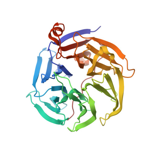
Human senescence marker protein 30 (SMP30), which functions enzymatically as a lactonase, hydrolyzes various carbohydrate lactones. The penultimate step in vitamin-C biosynthesis is catalyzed by this enzyme in nonprimate mammals. It has also been implicated as an organophosphate hydrolase, with the ability to hydrolyze diisopropyl phosphofluoridate and other nerve agents. SMP30 was originally identified as an aging marker protein, whose expression decreased androgen independently in aging cells. SMP30 is also referred to as regucalcin and has been suggested to have functions in calcium homeostasis. The crystal structure of the human enzyme has been solved from X-ray diffraction data collected to a resolution of 1.4 A. The protein has a 6-bladed beta-propeller fold, and it contains a single metal ion. Crystal structures have been solved with the metal site bound with either a Ca(2+) or a Zn(2+) atom. The catalytic role of the metal ion has been confirmed by mutagenesis of the metal coordinating residues. Kinetic studies using the substrate gluconolactone showed a k(cat) preference of divalent cations in the order Zn(2+) > Mn(2+) > Ca(2+) > Mg(2+). Notably, the Ca(2+) had a significantly higher value of K(d) compared to those of the other metal ions tested (566, 82, 7, and 0.6 mum for Ca(2+), Mg(2+), Zn(2+), and Mn(2+), respectively), suggesting that the Ca(2+)-bound form may be physiologically relevant for stressed cells with an elevated free calcium level.
Organizational Affiliation:
Department of Chemistry and Biochemistry, University of Delaware, Newark, Delaware 19716, USA.