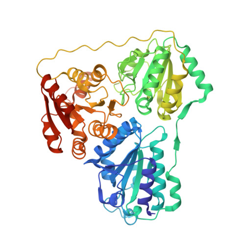
Structural and kinetic studies on native intermediates and an intermediate analogue in benzoylformate decarboxylase reveal a least motion mechanism with an unprecedented short-lived predecarboxylation intermediate.
Bruning, M., Berheide, M., Meyer, D., Golbik, R., Bartunik, H., Liese, A., Tittmann, K.(2009) Biochemistry 48: 3258-3268
- PubMed: 19182954
- DOI: https://doi.org/10.1021/bi801957d
- Primary Citation of Related Structures:
3FZN - PubMed Abstract:
The thiamin diphosphate- (ThDP-) dependent enzyme benzoylformate decarboxylase (BFDC) catalyzes the nonoxidative decarboxylation of benzoylformic acid to benzaldehyde and carbon dioxide. To date, no structural information for a cofactor-bound reaction intermediate in BFDC is available. For kinetic analysis, a chromophoric substrate analogue was employed that produces various absorbing intermediates during turnover but is a poor substrate with a 10(4)-fold compromised kcat. Here, we have analyzed the steady-state distribution of native intermediates by a combined chemical quench/1H NMR spectroscopic approach and estimated the net rate constants of elementary catalytic steps. At substrate saturation, carbonyl addition of the substrate to the cofactor (k' approximately 500 s-1 at 30 degrees C) and elimination of benzaldehyde (k' approximately 2.400 s-1) were found to be partially rate-determining for catalysis, whereas decarboxylation of the transient 2-mandelyl-ThDP intermediate is 1 order of magnitude faster with k' approximately 16.000 s-1, the largest rate constant of decarboxylation in any thiamin enzyme characterized so far. The X-ray structure of a predecarboxylation intermediate analogue was determined to 1.6 A after cocrystallization of BFDC from Pseudomonas putida with benzoylphosphonic acid methyl ester. In contrast to the free acid, for which irreversible phosphorylation of active center Ser26 was reported, the methyl ester forms a covalent adduct with ThDP with a similar configuration at C2alpha as observed for other thiamin enzymes. The C2-C2alpha bond of the intermediate analogue is out of plane by 7degrees, indicating strain. The phosphonate part of the adduct forms hydrogen bonds with Ser26 and His281, and the 1-OH group is held in place by interactions with His70 and the 4'-amino group of ThDP. The phenyl ring accommodates in a hydrophobic pocket formed by Phe464, Phe397, Leu109, and Leu403. A comparison with the previously determined structure of BFDC in noncovalent complex with the inhibitor (R)-mandelate suggests a least motion mechanism. Binding of benzoylphosphonic acid methyl ester to BFDC was further characterized by CD spectroscopy and stopped-flow kinetics, indicating a two-step binding mechanism with a 200-fold slower carbonyl addition to ThDP than determined for benzoylformic acid, in line with the observed slight structural reorganization of Phe464 due to steric clashes with the phosphonate moiety.
Organizational Affiliation:
Institute of Technical Biocatalysis, Hamburg UniVersity of Technology, Denickestrasse 15, D-21073 Hamburg, Germany.