3FYT

Crystal structure of Bacillus pumilus acetyl xylan esterase S181A mutant in complex with beta-D-xylopyranose


Experimental Data Snapshot

  • Method: X-RAY DIFFRACTION
  • Resolution: 2.58 Å
  • R-Value Free: 0.241 
  • R-Value Work: 0.188 
  • R-Value Observed: 0.188 

wwPDB Validation   3D Report Full Report


Ligand Structure Quality Assessment 


This is version 2.2 of the entry. See complete history


Literature

Structural and functional studies of Bacillus pumilus acetyl xylan esterase

Krastanova, I.Cassetta, A.Mastihubova, M.Biely, P.Lamba, D.

To be published.

Macromolecules
Find similar proteins by:  (by identity cutoff)  |  3D Structure
Entity ID: 1
MoleculeChains Sequence LengthOrganismDetailsImage
Acetyl xylan esterase320Bacillus pumilusMutation(s): 1 
Gene Names: axe
EC: 3.1.1.6
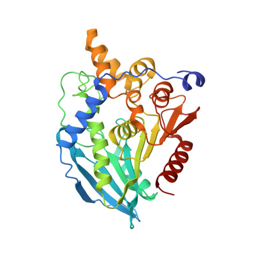
UniProt
Find proteins for Q9K5F2 (Bacillus pumilus)
Explore Q9K5F2 
Go to UniProtKB:  Q9K5F2
Entity Groups  
Sequence Clusters30% Identity50% Identity70% Identity90% Identity95% Identity100% Identity
UniProt GroupQ9K5F2
Sequence Annotations
Expand
  • Reference Sequence
Small Molecules
Ligands 2 Unique
IDChains Name / Formula / InChI Key2D Diagram3D Interactions
XYP
Query on XYP

Download Ideal Coordinates CCD File 
AA [auth E]
DA [auth F]
JA [auth H]
QA [auth L]
S [auth C]
AA [auth E],
DA [auth F],
JA [auth H],
QA [auth L],
S [auth C],
T [auth C]
beta-D-xylopyranose
C5 H10 O5
SRBFZHDQGSBBOR-KKQCNMDGSA-N
CL
Query on CL

Download Ideal Coordinates CCD File 
BA [auth E]
CA [auth E]
EA [auth F]
FA [auth F]
GA [auth F]
BA [auth E],
CA [auth E],
EA [auth F],
FA [auth F],
GA [auth F],
HA [auth G],
IA [auth G],
KA [auth H],
LA [auth H],
M [auth A],
MA [auth H],
N [auth A],
NA [auth I],
O [auth A],
OA [auth I],
P [auth B],
PA [auth I],
Q [auth B],
R [auth B],
RA [auth L],
SA [auth L],
TA [auth L],
U [auth C],
UA [auth M],
V [auth C],
VA [auth M],
W [auth C],
WA [auth M],
X [auth D],
XA [auth N],
Y [auth D],
YA [auth N],
Z [auth D],
ZA [auth N]
CHLORIDE ION
Cl
VEXZGXHMUGYJMC-UHFFFAOYSA-M
Experimental Data & Validation

Experimental Data

  • Method: X-RAY DIFFRACTION
  • Resolution: 2.58 Å
  • R-Value Free: 0.241 
  • R-Value Work: 0.188 
  • R-Value Observed: 0.188 
  • Space Group: P 1 21 1
Unit Cell:
Length ( Å )Angle ( ˚ )
a = 87.186α = 90
b = 167.986β = 91.17
c = 144.471γ = 90
Software Package:
Software NamePurpose
DENZOdata reduction
SCALEPACKdata scaling
AMoREphasing
CNSrefinement
PDB_EXTRACTdata extraction
ProDCdata collection
CNSphasing

Structure Validation

View Full Validation Report


Ligand Structure Quality Assessment 


Entry History 

Deposition Data

Revision History  (Full details and data files)

  • Version 1.0: 2010-01-26
    Type: Initial release
  • Version 1.1: 2011-07-13
    Changes: Version format compliance
  • Version 1.2: 2017-11-01
    Changes: Refinement description
  • Version 2.0: 2020-07-29
    Type: Remediation
    Reason: Carbohydrate remediation
    Changes: Atomic model, Data collection, Derived calculations, Structure summary
  • Version 2.1: 2021-10-20
    Changes: Database references, Structure summary
  • Version 2.2: 2023-09-06
    Changes: Data collection, Refinement description