3FYR

Crystal structure of the sporulation histidine kinase inhibitor Sda from Bacillus subtilis


Experimental Data Snapshot

  • Method: X-RAY DIFFRACTION
  • Resolution: 1.97 Å
  • R-Value Free: 0.304 
  • R-Value Work: 0.234 
  • R-Value Observed: 0.238 

wwPDB Validation   3D Report Full Report


This is version 1.2 of the entry. See complete history


Literature

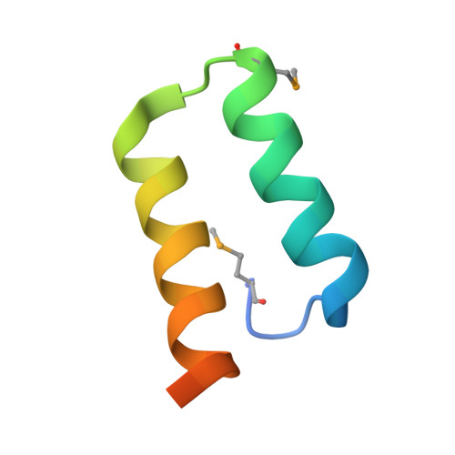
Structure of the sporulation histidine kinase inhibitor Sda from Bacillus subtilis and insights into its solution state

Jacques, D.A.Streamer, M.Rowland, S.L.King, G.F.Guss, J.M.Trewhella, J.Langley, D.B.

(2009) Acta Crystallogr D Biol Crystallogr 65: 574-581

  • DOI: https://doi.org/10.1107/S090744490901169X
  • Primary Citation of Related Structures:  
    3FYR

  • PubMed Abstract: 

    The crystal structure of the DNA-damage checkpoint inhibitor of sporulation, Sda, from Bacillus subtilis, has been solved by the MAD technique using selenomethionine-substituted protein. The structure closely resembles that previously solved by NMR, as well as the structure of a homologue from Geobacillus stearothermophilus solved in complex with the histidine kinase KinB. The structure contains three molecules in the asymmetric unit. The unusual trimeric arrangement, which lacks simple internal symmetry, appears to be preserved in solution based on an essentially ideal fit to previously acquired scattering data for Sda in solution. This interpretation contradicts previous findings that Sda was monomeric or dimeric in solution. This study demonstrates the difficulties that can be associated with the characterization of small proteins and the value of combining multiple biophysical techniques. It also emphasizes the importance of understanding the physical principles behind these techniques and therefore their limitations.


  • Organizational Affiliation

    School of Molecular and Microbial Biosciences, University of Sydney, Australia.


Macromolecules
Find similar proteins by:  (by identity cutoff)  |  3D Structure
Entity ID: 1
MoleculeChains Sequence LengthOrganismDetailsImage
Sporulation inhibitor sda
A, B, C
48Bacillus subtilis subsp. subtilis str. 168Mutation(s): 0 
Gene Names: BSU25690sda
UniProt
Find proteins for Q7WY62 (Bacillus subtilis (strain 168))
Explore Q7WY62 
Go to UniProtKB:  Q7WY62
Entity Groups  
Sequence Clusters30% Identity50% Identity70% Identity90% Identity95% Identity100% Identity
UniProt GroupQ7WY62
Sequence Annotations
Expand
  • Reference Sequence
Small Molecules
Modified Residues  1 Unique
IDChains TypeFormula2D DiagramParent
MSE
Query on MSE
A, B, C
L-PEPTIDE LINKINGC5 H11 N O2 SeMET
Experimental Data & Validation

Experimental Data

  • Method: X-RAY DIFFRACTION
  • Resolution: 1.97 Å
  • R-Value Free: 0.304 
  • R-Value Work: 0.234 
  • R-Value Observed: 0.238 
  • Space Group: P 41 21 2
Unit Cell:
Length ( Å )Angle ( ˚ )
a = 36.972α = 90
b = 36.972β = 90
c = 167.173γ = 90
Software Package:
Software NamePurpose
DENZOdata reduction
SCALEPACKdata scaling
SOLVEphasing
RESOLVEphasing
REFMACrefinement
PDB_EXTRACTdata extraction
HKL-2000data collection

Structure Validation

View Full Validation Report


Entry History 

Deposition Data

Revision History  (Full details and data files)

  • Version 1.0: 2009-06-23
    Type: Initial release
  • Version 1.1: 2011-07-13
    Changes: Version format compliance
  • Version 1.2: 2017-11-01
    Changes: Refinement description