3FNU

Crystal structure of KNI-10006 bound histo-aspartic protease (HAP) from Plasmodium falciparum


Experimental Data Snapshot

  • Method: X-RAY DIFFRACTION
  • Resolution: 3.00 Å
  • R-Value Free: 0.253 
  • R-Value Work: 0.222 
  • R-Value Observed: 0.223 

wwPDB Validation   3D Report Full Report


Ligand Structure Quality Assessment 


This is version 1.3 of the entry. See complete history


Literature

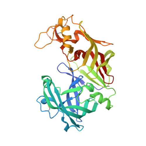
Crystal structures of the histo-aspartic protease (HAP) from Plasmodium falciparum.

Bhaumik, P.Xiao, H.Parr, C.L.Kiso, Y.Gustchina, A.Yada, R.Y.Wlodawer, A.

(2009) J Mol Biol 388: 520-540

  • DOI: https://doi.org/10.1016/j.jmb.2009.03.011
  • Primary Citation of Related Structures:  
    3FNS, 3FNT, 3FNU

  • PubMed Abstract: 

    The structures of recombinant histo-aspartic protease (HAP) from malaria-causing parasite Plasmodium falciparum as apoenzyme and in complex with two inhibitors, pepstatin A and KNI-10006, were solved at 2.5-, 3.3-, and 3.05-A resolutions, respectively. In the apoenzyme crystals, HAP forms a tight dimer not seen previously in any aspartic protease. The interactions between the monomers affect the conformation of two flexible loops, the functionally important "flap" (residues 70-83) and its structural equivalent in the C-terminal domain (residues 238-245), as well as the orientation of helix 225-235. The flap is found in an open conformation in the apoenzyme. Unexpectedly, the active site of the apoenzyme contains a zinc ion tightly bound to His32 and Asp215 from one monomer and to Glu278A from the other monomer, with the coordination of Zn resembling that seen in metalloproteases. The flap is closed in the structure of the pepstatin A complex, whereas it is open in the complex with KNI-10006. Although the binding mode of pepstatin A is significantly different from that in other pepsin-like aspartic proteases, its location in the active site makes unlikely the previously proposed hypothesis that HAP is a serine protease. The binding mode of KNI-10006 is unusual compared with the binding of other inhibitors from the KNI series to aspartic proteases. The novel features of the HAP active site could facilitate design of specific inhibitors used in the development of antimalarial drugs.


  • Organizational Affiliation

    Protein Structure Section, Macromolecular Crystallography Laboratory, National Cancer Institute, Frederick, MD 21702, USA.


Macromolecules
Find similar proteins by:  (by identity cutoff)  |  3D Structure
Entity ID: 1
MoleculeChains Sequence LengthOrganismDetailsImage
HAP protein
A, B, C, D
332Plasmodium falciparum 3D7Mutation(s): 0 
Gene Names: HAPPF14_0078
UniProt
Find proteins for Q8IM15 (Plasmodium falciparum (isolate 3D7))
Explore Q8IM15 
Go to UniProtKB:  Q8IM15
Entity Groups  
Sequence Clusters30% Identity50% Identity70% Identity90% Identity95% Identity100% Identity
UniProt GroupQ8IM15
Sequence Annotations
Expand
  • Reference Sequence
Small Molecules
Ligands 1 Unique
IDChains Name / Formula / InChI Key2D Diagram3D Interactions
006
Query on 006

Download Ideal Coordinates CCD File 
E [auth A],
F [auth B],
G [auth C],
H [auth D]
(4R)-3-[(2S,3S)-3-{[(2,6-dimethylphenoxy)acetyl]amino}-2-hydroxy-4-phenylbutanoyl]-N-[(1S,2R)-2-hydroxy-2,3-dihydro-1H-inden-1-yl]-5,5-dimethyl-1,3-thiazolidine-4-carboxamide
C35 H41 N3 O6 S
KKTYZYHUPKXLPL-RIQJEONASA-N
Binding Affinity Annotations 
IDSourceBinding Affinity
006 BindingDB:  3FNU IC50: 690 (nM) from 1 assay(s)
Experimental Data & Validation

Experimental Data

  • Method: X-RAY DIFFRACTION
  • Resolution: 3.00 Å
  • R-Value Free: 0.253 
  • R-Value Work: 0.222 
  • R-Value Observed: 0.223 
  • Space Group: I 41 2 2
Unit Cell:
Length ( Å )Angle ( ˚ )
a = 166.1α = 90
b = 166.1β = 90
c = 276.9γ = 90
Software Package:
Software NamePurpose
MAR345data collection
PHASERphasing
REFMACrefinement
XDSdata reduction
XDSdata scaling

Structure Validation

View Full Validation Report


Ligand Structure Quality Assessment 


Entry History 

Deposition Data

Revision History  (Full details and data files)

  • Version 1.0: 2009-05-12
    Type: Initial release
  • Version 1.1: 2011-07-13
    Changes: Advisory, Atomic model, Database references, Derived calculations, Non-polymer description, Source and taxonomy, Structure summary, Version format compliance
  • Version 1.2: 2013-02-27
    Changes: Other
  • Version 1.3: 2017-11-01
    Changes: Refinement description