3FMT

Crystal structure of SeqA bound to DNA


Experimental Data Snapshot

  • Method: X-RAY DIFFRACTION
  • Resolution: 2.98 Å
  • R-Value Free: 0.245 
  • R-Value Work: 0.218 
  • R-Value Observed: 0.219 

wwPDB Validation   3D Report Full Report


This is version 1.4 of the entry. See complete history


Literature

Structural insights into the cooperative binding of SeqA to a tandem GATC repeat

Chung, Y.S.Brendler, T.Austin, S.Guarne, A.

(2009) Nucleic Acids Res 37: 3143-3152

  • DOI: https://doi.org/10.1093/nar/gkp151
  • Primary Citation of Related Structures:  
    3FMT

  • PubMed Abstract: 

    SeqA is a negative regulator of DNA replication in Escherichia coli and related bacteria that functions by sequestering the origin of replication and facilitating its resetting after every initiation event. Inactivation of the seqA gene leads to unsynchronized rounds of replication, abnormal localization of nucleoids and increased negative superhelicity. Excess SeqA also disrupts replication synchrony and affects cell division. SeqA exerts its functions by binding clusters of transiently hemimethylated GATC sequences generated during replication. However, the molecular mechanisms that trigger formation and disassembly of such complex are unclear. We present here the crystal structure of a dimeric mutant of SeqA [SeqADelta(41-59)-A25R] bound to tandem hemimethylated GATC sites. The structure delineates how SeqA forms a high-affinity complex with DNA and it suggests why SeqA only recognizes GATC sites at certain spacings. The SeqA-DNA complex also unveils additional protein-protein interaction surfaces that mediate the formation of higher ordered complexes upon binding to newly replicated DNA. Based on this data, we propose a model describing how SeqA interacts with newly replicated DNA within the origin of replication and at the replication forks.


  • Organizational Affiliation

    Department of Biochemistry and Biomedical Sciences, Health Sciences Center, McMaster University, 1200 Main Street West, Hamilton, ON L8N 3Z5, Canada.


Macromolecules

Find similar proteins by:  (by identity cutoff)  |  3D Structure
Entity ID: 1
MoleculeChains Sequence LengthOrganismDetailsImage
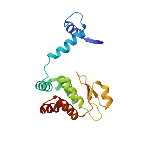
Protein seqA
A, B, E, F
162Escherichia coli K-12Mutation(s): 1 
Gene Names: b0687JW0674seqA
UniProt
Find proteins for P0AFY8 (Escherichia coli (strain K12))
Explore P0AFY8 
Go to UniProtKB:  P0AFY8
Entity Groups  
Sequence Clusters30% Identity50% Identity70% Identity90% Identity95% Identity100% Identity
UniProt GroupP0AFY8
Sequence Annotations
Expand
  • Reference Sequence

Find similar nucleic acids by:  Sequence   |   3D Structure  

Entity ID: 2
MoleculeChains LengthOrganismImage
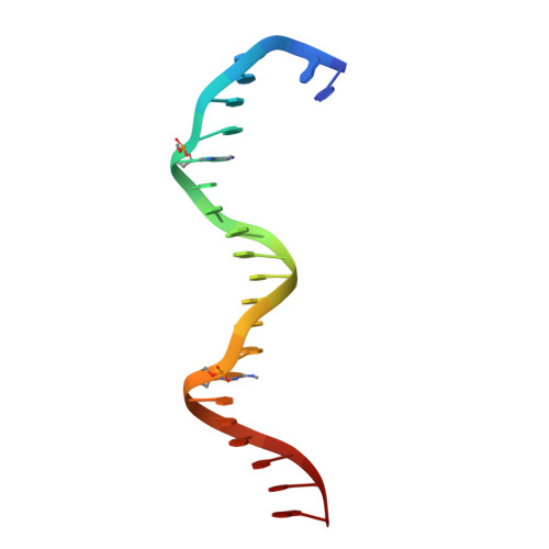
5'-D(*GP*AP*GP*TP*CP*GP*(6MA)P*TP*CP*GP*GP*CP*GP*GP*GP*(6MA)P*TP*CP*CP*TP*TP*A)-3'
C, G
22N/A
Sequence Annotations
Expand
  • Reference Sequence

Find similar nucleic acids by:  Sequence   |   3D Structure  

Entity ID: 3
MoleculeChains LengthOrganismImage
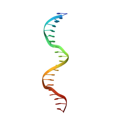
5'-D(*TP*CP*TP*AP*AP*GP*GP*AP*TP*CP*CP*CP*GP*CP*CP*GP*AP*TP*CP*GP*AP*C)-3'
D, H
22N/A
Sequence Annotations
Expand
  • Reference Sequence
Experimental Data & Validation

Experimental Data

  • Method: X-RAY DIFFRACTION
  • Resolution: 2.98 Å
  • R-Value Free: 0.245 
  • R-Value Work: 0.218 
  • R-Value Observed: 0.219 
  • Space Group: H 3
Unit Cell:
Length ( Å )Angle ( ˚ )
a = 121.839α = 90
b = 121.839β = 90
c = 279.133γ = 120
Software Package:
Software NamePurpose
CBASSdata collection
PHASERphasing
PHENIXrefinement
HKL-2000data reduction
HKL-2000data scaling

Structure Validation

View Full Validation Report


Entry History 

Deposition Data

Revision History  (Full details and data files)

  • Version 1.0: 2009-04-28
    Type: Initial release
  • Version 1.1: 2011-07-13
    Changes: Version format compliance
  • Version 1.2: 2017-07-26
    Changes: Refinement description, Source and taxonomy
  • Version 1.3: 2021-10-20
    Changes: Database references, Derived calculations
  • Version 1.4: 2023-09-06
    Changes: Data collection, Refinement description