3FLK

Crystal Structure of Tartrate Dehydrogenase from Pseudomonas putida in complex with NADH, oxalate and metal ion


Experimental Data Snapshot

  • Method: X-RAY DIFFRACTION
  • Resolution: 2.00 Å
  • R-Value Free: 0.227 
  • R-Value Work: 0.208 
  • R-Value Observed: 0.209 

wwPDB Validation   3D Report Full Report


Ligand Structure Quality Assessment 


This is version 1.3 of the entry. See complete history


Literature

Structural characterization of tartrate dehydrogenase: a versatile enzyme catalyzing multiple reactions.

Malik, R.Viola, R.E.

(2010) Acta Crystallogr D Biol Crystallogr 66: 673-684

  • DOI: https://doi.org/10.1107/S0907444910008851
  • Primary Citation of Related Structures:  
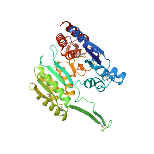
    3FLK

  • PubMed Abstract: 

    The first structure of an NAD-dependent tartrate dehydrogenase (TDH) has been solved to 2 A resolution by single anomalous diffraction (SAD) phasing as a complex with the intermediate analog oxalate, Mg(2+) and NADH. This TDH structure from Pseudomonas putida has a similar overall fold and domain organization to other structurally characterized members of the hydroxy-acid dehydrogenase family. However, there are considerable differences between TDH and these functionally related enzymes in the regions connecting the core secondary structure and in the relative positioning of important loops and helices. The active site in these complexes is highly ordered, allowing the identification of the substrate-binding and cofactor-binding groups and the ligands to the metal ions. Residues from the adjacent subunit are involved in both the substrate and divalent metal ion binding sites, establishing a dimer as the functional unit and providing structural support for an alternating-site reaction mechanism. The divalent metal ion plays a prominent role in substrate binding and orientation, together with several active-site arginines. Functional groups from both subunits form the cofactor-binding site and the ammonium ion aids in the orientation of the nicotinamide ring of the cofactor. A lysyl amino group (Lys192) is the base responsible for the water-mediated proton abstraction from the C2 hydroxyl group of the substrate that begins the catalytic reaction, followed by hydride transfer to NAD. A tyrosyl hydroxyl group (Tyr141) functions as a general acid to protonate the enolate intermediate. Each substrate undergoes the initial hydride transfer, but differences in substrate orientation are proposed to account for the different reactions catalyzed by TDH.


  • Organizational Affiliation

    Department of Chemistry, University of Toledo, Toledo, Ohio 43606, USA.


Macromolecules
Find similar proteins by:  (by identity cutoff)  |  3D Structure
Entity ID: 1
MoleculeChains Sequence LengthOrganismDetailsImage
Tartrate dehydrogenase/decarboxylase
A, B, C, D
364Pseudomonas putidaMutation(s): 1 
EC: 1.1.1.93
UniProt
Find proteins for Q51945 (Pseudomonas putida)
Explore Q51945 
Go to UniProtKB:  Q51945
Entity Groups  
Sequence Clusters30% Identity50% Identity70% Identity90% Identity95% Identity100% Identity
UniProt GroupQ51945
Sequence Annotations
Expand
  • Reference Sequence
Small Molecules
Ligands 6 Unique
IDChains Name / Formula / InChI Key2D Diagram3D Interactions
NAI
Query on NAI

Download Ideal Coordinates CCD File 
DA [auth D],
F [auth A],
N [auth B],
V [auth C]
1,4-DIHYDRONICOTINAMIDE ADENINE DINUCLEOTIDE
C21 H29 N7 O14 P2
BOPGDPNILDQYTO-NNYOXOHSSA-N
DTT
Query on DTT

Download Ideal Coordinates CCD File 
CA [auth C],
KA [auth D],
T [auth B]
2,3-DIHYDROXY-1,4-DITHIOBUTANE
C4 H10 O2 S2
VHJLVAABSRFDPM-IMJSIDKUSA-N
SO4
Query on SO4

Download Ideal Coordinates CCD File 
AA [auth C]
BA [auth C]
GA [auth D]
HA [auth D]
IA [auth D]
AA [auth C],
BA [auth C],
GA [auth D],
HA [auth D],
IA [auth D],
J [auth A],
JA [auth D],
K [auth A],
L [auth A],
M [auth A],
Q [auth B],
R [auth B],
S [auth B],
Z [auth C]
SULFATE ION
O4 S
QAOWNCQODCNURD-UHFFFAOYSA-L
OXL
Query on OXL

Download Ideal Coordinates CCD File 
EA [auth D],
G [auth A],
O [auth B],
W [auth C]
OXALATE ION
C2 O4
MUBZPKHOEPUJKR-UHFFFAOYSA-L
MG
Query on MG

Download Ideal Coordinates CCD File 
E [auth A],
I [auth A],
U [auth C],
Y [auth C]
MAGNESIUM ION
Mg
JLVVSXFLKOJNIY-UHFFFAOYSA-N
NH4
Query on NH4

Download Ideal Coordinates CCD File 
FA [auth D],
H [auth A],
P [auth B],
X [auth C]
AMMONIUM ION
H4 N
QGZKDVFQNNGYKY-UHFFFAOYSA-O
Modified Residues  1 Unique
IDChains TypeFormula2D DiagramParent
MSE
Query on MSE
A, B, C, D
L-PEPTIDE LINKINGC5 H11 N O2 SeMET
Experimental Data & Validation

Experimental Data

  • Method: X-RAY DIFFRACTION
  • Resolution: 2.00 Å
  • R-Value Free: 0.227 
  • R-Value Work: 0.208 
  • R-Value Observed: 0.209 
  • Space Group: P 43 21 2
Unit Cell:
Length ( Å )Angle ( ˚ )
a = 117.278α = 90
b = 117.278β = 90
c = 291.324γ = 90
Software Package:
Software NamePurpose
HKL-2000data collection
SHARPphasing
REFMACrefinement
HKL-2000data reduction
HKL-2000data scaling

Structure Validation

View Full Validation Report


Ligand Structure Quality Assessment 


Entry History 

Deposition Data

Revision History  (Full details and data files)

  • Version 1.0: 2009-12-22
    Type: Initial release
  • Version 1.1: 2011-07-13
    Changes: Version format compliance
  • Version 1.2: 2013-10-30
    Changes: Non-polymer description
  • Version 1.3: 2021-10-20
    Changes: Database references, Derived calculations