3FF5

Crystal structure of the conserved N-terminal domain of the peroxisomal matrix-protein-import receptor, Pex14p


Experimental Data Snapshot

  • Method: X-RAY DIFFRACTION
  • Resolution: 1.80 Å
  • R-Value Free: 0.211 
  • R-Value Work: 0.179 
  • R-Value Observed: 0.181 

wwPDB Validation   3D Report Full Report


Ligand Structure Quality Assessment 


This is version 1.3 of the entry. See complete history


Literature

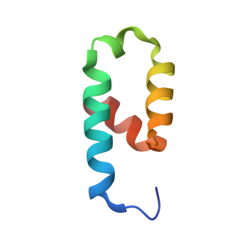
Crystal structure of the conserved N-terminal domain of the peroxisomal matrix protein import receptor, Pex14p

Su, J.R.Takeda, K.Tamura, S.Fujiki, Y.Miki, K.

(2009) Proc Natl Acad Sci U S A 106: 417-421

  • DOI: https://doi.org/10.1073/pnas.0808681106
  • Primary Citation of Related Structures:  
    3FF5

  • PubMed Abstract: 

    Pex14p is a central component of the peroxisomal protein import machinery, in which the conserved N-terminal domain mediates dynamic interactions with other peroxins including Pex5p, Pex13p, and Pex19p. Here, we report the crystal structure of the conserved N-terminal domain of Pex14p with a three-helix bundle. A hydrophobic surface is composed of the conserved residues, of which two phenylalanine residues (Phe-35 and Phe-52) protrude to the solvent. Consequently, two putative binding pockets suitable for recognizing the helical WXXXF/Y motif of Pex5p are formed on the surface by the two phenylalanine residues accompanying with positively charged residues. The structural feature agrees well with our earlier findings where F35A/L36A and F52A/L53A mutants were impaired in the interactions with other peroxins such as Pex5p and Pex13p. Pex14p variants each with Phe-to-Ala mutation at positions 35, 52, and 35/52, respectively, were defective in restoring the impaired peroxisomal protein import in pex14 Chinese hamster ovary mutant ZP161 cells. Moreover, in GST pull-down assays His(6)-Pex5pL bound only to GST-Pex14p(25-70), not to any of GST-Pex14p(25-70)F35A, GST-Pex14p(25-70)F52A, and GST-Pex14p(25-70)F35A/F52A. Endogenous Pex5p was recruited to FLAG-Pex14p on peroxisomes in vivo but barely to FLAG-Pex14pF35A, FLAG-Pex14pF52A, and FLAG-Pex14pF35A/F52A. Collectively, Phe-35 and Phe-52 are essential for the Pex14p functions, including the interaction between Pex14p and Pex5p.


  • Organizational Affiliation

    Department of Chemistry, Graduate School of Science, Kyoto University, Sakyo-ku, Kyoto 606-8502, Japan.


Macromolecules
Find similar proteins by:  (by identity cutoff)  |  3D Structure
Entity ID: 1
MoleculeChains Sequence LengthOrganismDetailsImage
Peroxisomal biogenesis factor 14
A, B
54Rattus norvegicusMutation(s): 0 
Gene Names: Pex14
UniProt
Find proteins for Q642G4 (Rattus norvegicus)
Explore Q642G4 
Go to UniProtKB:  Q642G4
Entity Groups  
Sequence Clusters30% Identity50% Identity70% Identity90% Identity95% Identity100% Identity
UniProt GroupQ642G4
Sequence Annotations
Expand
  • Reference Sequence
Small Molecules
Ligands 1 Unique
IDChains Name / Formula / InChI Key2D Diagram3D Interactions
DPW
Query on DPW

Download Ideal Coordinates CCD File 
C [auth A]decyl 2-trimethylazaniumylethyl phosphate
C15 H34 N O4 P
VVVDGSCGBGBGFN-UHFFFAOYSA-N
Experimental Data & Validation

Experimental Data

  • Method: X-RAY DIFFRACTION
  • Resolution: 1.80 Å
  • R-Value Free: 0.211 
  • R-Value Work: 0.179 
  • R-Value Observed: 0.181 
  • Space Group: I 2 3
Unit Cell:
Length ( Å )Angle ( ˚ )
a = 90.57α = 90
b = 90.57β = 90
c = 90.57γ = 90
Software Package:
Software NamePurpose
SOLVEphasing
REFMACrefinement
HKL-2000data reduction
HKL-2000data scaling

Structure Validation

View Full Validation Report


Ligand Structure Quality Assessment 


Entry History 

Deposition Data

Revision History  (Full details and data files)

  • Version 1.0: 2008-12-30
    Type: Initial release
  • Version 1.1: 2011-07-13
    Changes: Version format compliance
  • Version 1.2: 2012-05-02
    Changes: Database references
  • Version 1.3: 2023-12-27
    Changes: Data collection, Database references, Derived calculations