3F86

An alpha/beta-Peptide Helix Bundle with a Pure beta-Amino Acid Core and a Distinctive Quaternary Structure: GCN4pLI derivative with beta residues at a and d heptad positions


Experimental Data Snapshot

  • Method: X-RAY DIFFRACTION
  • Resolution: 2.00 Å
  • R-Value Free: 0.266 
  • R-Value Work: 0.200 
  • R-Value Observed: 0.203 

wwPDB Validation   3D Report Full Report


This is version 2.0 of the entry. See complete history


Literature

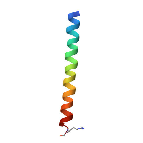
An alpha/beta-peptide helix bundle with a pure beta3-amino acid core and a distinctive quaternary structure.

Giuliano, M.W.Horne, W.S.Gellman, S.H.

(2009) J Am Chem Soc 131: 9860-9861

  • DOI: https://doi.org/10.1021/ja8099294
  • Primary Citation of Related Structures:  
    3F86, 3F87

  • PubMed Abstract: 

    Helix bundles are among the most widely studied tertiary and quaternary structural motifs in proteins. Here we present the crystal structure of an alpha/beta-peptide foldamer that adopts a tetrameric helix-bundle quaternary structure with a hydrophobic core composed solely of beta-amino acids. The structure displays features that are unprecedented among all known helix bundles composed of either alpha-peptides or peptidic foldamers. The tetramer is characterized by an asymmetry of interaction between neighboring helices, and the side-chain packing within the hydrophobic core differs fundamentally from the knobs-into-holes arrangement typical of most helix bundles.


  • Organizational Affiliation

    Department of Chemistry, University of Wisconsin, Madison, Wisconsin 53706, USA.


Macromolecules
Find similar proteins by:  (by identity cutoff)  |  3D Structure
Entity ID: 1
MoleculeChains Sequence LengthOrganismDetailsImage
GCN4pLI-betaAD
A, B, C, D, E
A, B, C, D, E, F, G, H
34N/AMutation(s): 0 
Entity Groups  
Sequence Clusters30% Identity50% Identity70% Identity90% Identity95% Identity100% Identity
Sequence Annotations
Expand
  • Reference Sequence
Small Molecules
Modified Residues  1 Unique
IDChains TypeFormula2D DiagramParent
HMR
Query on HMR
A, B, C, D, E
A, B, C, D, E, F, G, H
L-PEPTIDE LINKINGC7 H16 N4 O2ARG
Experimental Data & Validation

Experimental Data

  • Method: X-RAY DIFFRACTION
  • Resolution: 2.00 Å
  • R-Value Free: 0.266 
  • R-Value Work: 0.200 
  • R-Value Observed: 0.203 
  • Space Group: P 1 21 1
Unit Cell:
Length ( Å )Angle ( ˚ )
a = 38.381α = 90
b = 68.72β = 100.93
c = 48.297γ = 90
Software Package:
Software NamePurpose
REFMACrefinement
PROTEUM PLUSdata collection
PROTEUM PLUSdata reduction
PROTEUM PLUSdata scaling
PHASERphasing

Structure Validation

View Full Validation Report


Entry History 

Deposition Data

Revision History  (Full details and data files)

  • Version 1.0: 2009-07-21
    Type: Initial release
  • Version 1.1: 2011-07-13
    Changes: Version format compliance
  • Version 2.0: 2023-11-15
    Changes: Atomic model, Data collection, Database references, Derived calculations