3F59

Crystal structure of ZU5-ANK, the spectrin binding region of human erythroid ankyrin


Experimental Data Snapshot

  • Method: X-RAY DIFFRACTION
  • Resolution: 2.00 Å
  • R-Value Free: 0.264 
  • R-Value Work: 0.216 
  • R-Value Observed: 0.218 

wwPDB Validation   3D Report Full Report


This is version 1.3 of the entry. See complete history


Literature

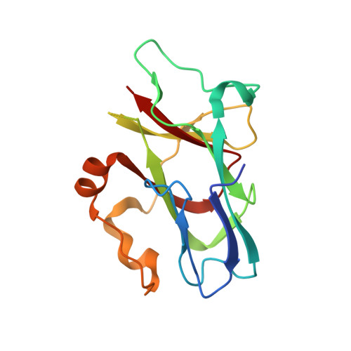
Structures of the spectrin-ankyrin interaction binding domains.

Ipsaro, J.J.Huang, L.Mondragon, A.

(2009) Blood 113: 5385-5393

  • DOI: https://doi.org/10.1182/blood-2008-10-184358
  • Primary Citation of Related Structures:  
    3F57, 3F59

  • PubMed Abstract: 

    As key components of the erythrocyte membrane skeleton, spectrin and ankyrin specifically interact to tether the spectrin cytoskeleton to the cell membrane. The structure of the spectrin binding domain of ankyrin and the ankyrin binding domain of spectrin have been solved to elucidate the structural basis for ankyrin-spectrin recognition. The structure of repeats 14 and 15 of spectrin shows that these repeats are similar to all other spectrin repeats. One feature that could account for the preference of ankyrin for these repeats is the presence of a conserved, negatively charged patch on one side of repeat 14. The structure of the ankyrin ZU5 domain shows a novel structure containing a beta core. The structure reveals that the canonical ZU5 consensus sequence is likely to be missing an important region that codes for a beta strand that forms part of the core of the domain. In addition, a positively charged region is suggestive of a binding surface for the negatively charged spectrin repeat 14. Previously reported mutants of ankyrin that map to this region lie mostly on the surface of the protein, although at least one is likely to be part of the core.


  • Organizational Affiliation

    Department of Biochemistry, Molecular Biology, and Cell Biology, Northwestern University, Evanston, IL 60208, USA.


Macromolecules
Find similar proteins by:  (by identity cutoff)  |  3D Structure
Entity ID: 1
MoleculeChains Sequence LengthOrganismDetailsImage
Ankyrin-1
A, B, C, D
161Homo sapiensMutation(s): 0 
Gene Names: ANK1ANK
UniProt & NIH Common Fund Data Resources
Find proteins for P16157 (Homo sapiens)
Explore P16157 
Go to UniProtKB:  P16157
PHAROS:  P16157
GTEx:  ENSG00000029534 
Entity Groups  
Sequence Clusters30% Identity50% Identity70% Identity90% Identity95% Identity100% Identity
UniProt GroupP16157
Sequence Annotations
Expand
  • Reference Sequence
Small Molecules
Ligands 1 Unique
IDChains Name / Formula / InChI Key2D Diagram3D Interactions
BR
Query on BR

Download Ideal Coordinates CCD File 
E [auth A]
F [auth A]
G [auth A]
H [auth A]
I [auth A]
E [auth A],
F [auth A],
G [auth A],
H [auth A],
I [auth A],
J [auth A],
K [auth B],
L [auth B],
M [auth B],
N [auth C],
O [auth C],
P [auth C],
Q [auth C],
R [auth D],
S [auth D],
T [auth D],
U [auth D],
V [auth D]
BROMIDE ION
Br
CPELXLSAUQHCOX-UHFFFAOYSA-M
Experimental Data & Validation

Experimental Data

  • Method: X-RAY DIFFRACTION
  • Resolution: 2.00 Å
  • R-Value Free: 0.264 
  • R-Value Work: 0.216 
  • R-Value Observed: 0.218 
  • Space Group: P 1 21 1
Unit Cell:
Length ( Å )Angle ( ˚ )
a = 38.04α = 90
b = 204.11β = 113.14
c = 43.07γ = 90
Software Package:
Software NamePurpose
MAR345data collection
SHARPphasing
REFMACrefinement
XDSdata reduction
SCALAdata scaling

Structure Validation

View Full Validation Report


Entry History 

Deposition Data

Revision History  (Full details and data files)

  • Version 1.0: 2009-02-24
    Type: Initial release
  • Version 1.1: 2011-07-13
    Changes: Advisory, Version format compliance
  • Version 1.2: 2017-10-25
    Changes: Refinement description
  • Version 1.3: 2023-12-27
    Changes: Data collection, Database references, Derived calculations