3EK7

Calcium-saturated GCaMP2 dimer


Experimental Data Snapshot

  • Method: X-RAY DIFFRACTION
  • Resolution: 1.85 Å
  • R-Value Free: 0.241 
  • R-Value Work: 0.189 
  • R-Value Observed: 0.191 

wwPDB Validation   3D Report Full Report


This is version 1.6 of the entry. See complete history


Literature

Crystal Structures of the GCaMP Calcium Sensor Reveal the Mechanism of Fluorescence Signal Change and Aid Rational Design

Akerboom, J.Rivera, J.D.Guilbe, M.M.Malave, E.C.Hernandez, H.H.Tian, L.Hires, S.A.Marvin, J.S.Looger, L.L.Schreiter, E.R.

(2009) J Biol Chem 284: 6455-6464

  • DOI: https://doi.org/10.1074/jbc.M807657200
  • Primary Citation of Related Structures:  
    3EK4, 3EK7, 3EK8, 3EKH, 3EKJ

  • PubMed Abstract: 

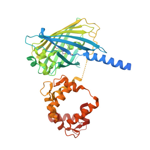
    The genetically encoded calcium indicator GCaMP2 shows promise for neural network activity imaging, but is currently limited by low signal-to-noise ratio. We describe x-ray crystal structures as well as solution biophysical and spectroscopic characterization of GCaMP2 in the calcium-free dark state, and in two calcium-bound bright states: a monomeric form that dominates at intracellular concentrations observed during imaging experiments and an unexpected domain-swapped dimer with decreased fluorescence. This series of structures provides insight into the mechanism of Ca2+-induced fluorescence change. Upon calcium binding, the calmodulin (CaM) domain wraps around the M13 peptide, creating a new domain interface between CaM and the circularly permuted enhanced green fluorescent protein domain. Residues from CaM alter the chemical environment of the circularly permuted enhanced green fluorescent protein chromophore and, together with flexible inter-domain linkers, block solvent access to the chromophore. Guided by the crystal structures, we engineered a series of GCaMP2 point mutants to probe the mechanism of GCaMP2 function and characterized one mutant with significantly improved signal-to-noise. The mutation is located at a domain interface and its effect on sensor function could not have been predicted in the absence of structural data.


  • Organizational Affiliation

    Janelia Farm Research Campus, Howard Hughes Medical Institute, Ashburn, Virginia 20147, USA.


Macromolecules
Find similar proteins by:  (by identity cutoff)  |  3D Structure
Entity ID: 1
MoleculeChains Sequence LengthOrganismDetailsImage
Myosin light chain kinase, Green fluorescent protein, Calmodulin chimera449synthetic constructAequorea victoriaRattus norvegicusMutation(s): 1 
UniProt
Find proteins for P42212 (Aequorea victoria)
Explore P42212 
Go to UniProtKB:  P42212
Find proteins for P11799 (Gallus gallus)
Explore P11799 
Go to UniProtKB:  P11799
Find proteins for P0DP29 (Rattus norvegicus)
Explore P0DP29 
Go to UniProtKB:  P0DP29
Entity Groups  
Sequence Clusters30% Identity50% Identity70% Identity90% Identity95% Identity100% Identity
UniProt GroupsP42212P0DP29P11799
Sequence Annotations
Expand
  • Reference Sequence
Experimental Data & Validation

Experimental Data

  • Method: X-RAY DIFFRACTION
  • Resolution: 1.85 Å
  • R-Value Free: 0.241 
  • R-Value Work: 0.189 
  • R-Value Observed: 0.191 
  • Space Group: C 1 2 1
Unit Cell:
Length ( Å )Angle ( ˚ )
a = 126.131α = 90
b = 47.31β = 100.48
c = 68.94γ = 90
Software Package:
Software NamePurpose
SCALAdata scaling
PHASERphasing
REFMACrefinement
PDB_EXTRACTdata extraction
DENZOdata reduction
SCALEPACKdata scaling

Structure Validation

View Full Validation Report


Entry History 

Deposition Data

Revision History  (Full details and data files)

  • Version 1.0: 2008-12-16
    Type: Initial release
  • Version 1.1: 2011-07-13
    Changes: Version format compliance
  • Version 1.2: 2012-10-31
    Changes: Database references
  • Version 1.3: 2017-06-21
    Changes: Database references, Source and taxonomy
  • Version 1.4: 2017-10-25
    Changes: Refinement description
  • Version 1.5: 2023-08-30
    Changes: Data collection, Database references, Derived calculations, Refinement description
  • Version 1.6: 2023-11-15
    Changes: Data collection