3E4G

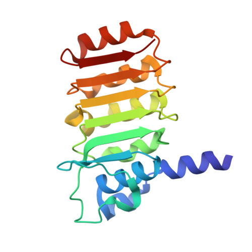
Crystal structure of bovine coupling Factor B, G28E mutant


Experimental Data Snapshot

  • Method: X-RAY DIFFRACTION
  • Resolution: 0.96 Å
  • R-Value Free: 0.164 
  • R-Value Work: 0.143 
  • R-Value Observed: 0.144 

wwPDB Validation   3D Report Full Report


This is version 1.3 of the entry. See complete history


Literature

Crystal structure of bovine mitochondrial factor B at 0.96-A resolution.

Lee, J.K.Belogrudov, G.I.Stroud, R.M.

(2008) Proc Natl Acad Sci U S A 105: 13379-13384

  • DOI: https://doi.org/10.1073/pnas.0805689105
  • Primary Citation of Related Structures:  
    3DZE, 3E2J, 3E3Z, 3E4G

  • PubMed Abstract: 

    Coupling factor B (FB) is a mitochondrial inner membrane polypeptide that facilitates the energy-driven catalysis of ATP synthesis in animal mitochondria by blocking a proton leak across the membrane. Here, we report the crystal structure of the bovine mitochondrial FB mutant with Gly-3-Glu substitution determined at a resolution of 0.96 A and that of the WT polypeptide at a resolution of 2.9 A. The structure reveals an oblong, oval-shaped molecule with a unique globular N-terminal domain that is proposed to be the membrane anchor domain and the capping region to the C-terminal leucine-rich repeats domain. A short N-terminal alpha-helix, which extends away from the molecule's body, is suggestive of functioning as an anchor for FB to the matrix side of the mitochondrial inner membrane. Identification of a bound Mg(2+) ion reveals that FB is a metalloprotein. We also report the cocrystal structures of FB bound with phenylarsine oxide and Cd(2+), two known inhibitors of the FB coupling activity.


  • Organizational Affiliation

    Department of Biochemistry and Biophysics, University of California, San Francisco, CA 94158-2517, USA.


Macromolecules
Find similar proteins by:  (by identity cutoff)  |  3D Structure
Entity ID: 1
MoleculeChains Sequence LengthOrganismDetailsImage
ATP synthase subunit s, mitochondrial176Bos taurusMutation(s): 1 
Gene Names: ATP5SATPW
EC: 3.6.3.14
UniProt
Find proteins for P22027 (Bos taurus)
Explore P22027 
Go to UniProtKB:  P22027
Entity Groups  
Sequence Clusters30% Identity50% Identity70% Identity90% Identity95% Identity100% Identity
UniProt GroupP22027
Sequence Annotations
Expand
  • Reference Sequence
Small Molecules
Ligands 2 Unique
IDChains Name / Formula / InChI Key2D Diagram3D Interactions
TRS
Query on TRS

Download Ideal Coordinates CCD File 
C [auth A]2-AMINO-2-HYDROXYMETHYL-PROPANE-1,3-DIOL
C4 H12 N O3
LENZDBCJOHFCAS-UHFFFAOYSA-O
MG
Query on MG

Download Ideal Coordinates CCD File 
B [auth A]MAGNESIUM ION
Mg
JLVVSXFLKOJNIY-UHFFFAOYSA-N
Experimental Data & Validation

Experimental Data

  • Method: X-RAY DIFFRACTION
  • Resolution: 0.96 Å
  • R-Value Free: 0.164 
  • R-Value Work: 0.143 
  • R-Value Observed: 0.144 
  • Space Group: C 1 2 1
Unit Cell:
Length ( Å )Angle ( ˚ )
a = 107.476α = 90
b = 50.01β = 109.37
c = 36.886γ = 90
Software Package:
Software NamePurpose
MOSFLMdata reduction
SCALAdata scaling
PHASERphasing
REFMACrefinement
PDB_EXTRACTdata extraction
Blu-Icedata collection

Structure Validation

View Full Validation Report


Entry History 

Deposition Data

Revision History  (Full details and data files)

  • Version 1.0: 2008-08-26
    Type: Initial release
  • Version 1.1: 2011-07-13
    Changes: Version format compliance
  • Version 1.2: 2021-10-20
    Changes: Database references, Derived calculations
  • Version 1.3: 2023-08-30
    Changes: Data collection, Refinement description