3D8N

Uroporphyrinogen III Synthase-Uroporphyringen III Complex


Experimental Data Snapshot

  • Method: X-RAY DIFFRACTION
  • Resolution: 1.90 Å
  • R-Value Free: 0.245 
  • R-Value Work: 0.206 
  • R-Value Observed: 0.208 

wwPDB Validation   3D Report Full Report


Ligand Structure Quality Assessment 


This is version 1.3 of the entry. See complete history


Literature

Structure and mechanistic implications of a uroporphyrinogen III synthase-product complex.

Schubert, H.L.Phillips, J.D.Heroux, A.Hill, C.P.

(2008) Biochemistry 47: 8648-8655

  • DOI: https://doi.org/10.1021/bi800635y
  • Primary Citation of Related Structures:  
    3D8N, 3D8R, 3D8S, 3D8T

  • PubMed Abstract: 

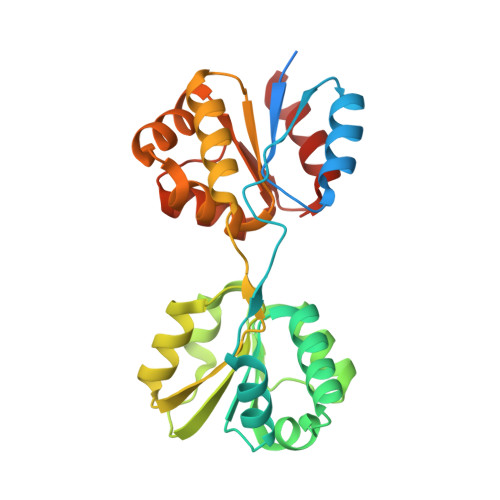
    Uroporphyrinogen III synthase (U3S) catalyzes the asymmetrical cyclization of a linear tetrapyrrole to form the physiologically relevant uroporphyrinogen III (uro'gen III) isomer during heme biosynthesis. Here, we report four apoenzyme and one product complex crystal structures of the Thermus thermophilus (HB27) U3S protein. The overlay of eight crystallographically unique U3S molecules reveals a huge range of conformational flexibility, including a "closed" product complex. The product, uro'gen III, binds between the two domains and is held in place by a network of hydrogen bonds between the product's side chain carboxylates and the protein's main chain amides. Interactions of the product A and B ring carboxylate side chains with both structural domains of U3S appear to dictate the relative orientation of the domains in the closed enzyme conformation and likely remain intact during catalysis. The product C and D rings are less constrained in the structure, consistent with the conformational changes required for the catalytic cyclization with inversion of D ring orientation. A conserved tyrosine residue is potentially positioned to facilitate loss of a hydroxyl from the substrate to initiate the catalytic reaction.


  • Organizational Affiliation

    Departments of Biochemistry and Internal Medicine, School of Medicine, University of Utah, Salt Lake City, Utah 84112, , USA. heidi@biochem.utah.edu


Macromolecules
Find similar proteins by:  (by identity cutoff)  |  3D Structure
Entity ID: 1
MoleculeChains Sequence LengthOrganismDetailsImage
Uroporphyrinogen-III synthase286Thermus thermophilus HB27Mutation(s): 0 
Gene Names: TT_C0312
EC: 4.2.1.75
UniProt
Find proteins for Q72KM1 (Thermus thermophilus (strain ATCC BAA-163 / DSM 7039 / HB27))
Explore Q72KM1 
Go to UniProtKB:  Q72KM1
Entity Groups  
Sequence Clusters30% Identity50% Identity70% Identity90% Identity95% Identity100% Identity
UniProt GroupQ72KM1
Sequence Annotations
Expand
  • Reference Sequence
Small Molecules
Ligands 1 Unique
IDChains Name / Formula / InChI Key2D Diagram3D Interactions
UP3
Query on UP3

Download Ideal Coordinates CCD File 
B [auth A]3,3',3'',3'''-[3,8,13,17-tetrakis(carboxymethyl)porphyrin-2,7,12,18-tetrayl]tetrapropanoic acid
C40 H38 N4 O16
VZVFNUAIRVUCEW-XFMUOGEMSA-N
Experimental Data & Validation

Experimental Data

  • Method: X-RAY DIFFRACTION
  • Resolution: 1.90 Å
  • R-Value Free: 0.245 
  • R-Value Work: 0.206 
  • R-Value Observed: 0.208 
  • Space Group: P 1 21 1
Unit Cell:
Length ( Å )Angle ( ˚ )
a = 35.743α = 90
b = 66.478β = 91.76
c = 55.459γ = 90
Software Package:
Software NamePurpose
DENZOdata reduction
SCALEPACKdata scaling
PHASERphasing
REFMACrefinement
PDB_EXTRACTdata extraction
ADSCdata collection
HKL-2000data reduction
HKL-2000data scaling

Structure Validation

View Full Validation Report


Ligand Structure Quality Assessment 


Entry History 

Deposition Data

Revision History  (Full details and data files)

  • Version 1.0: 2008-08-12
    Type: Initial release
  • Version 1.1: 2011-07-13
    Changes: Source and taxonomy, Version format compliance
  • Version 1.2: 2017-10-25
    Changes: Refinement description
  • Version 1.3: 2024-02-21
    Changes: Data collection, Database references, Derived calculations