Crystal structure of Mycobacterium tuberculosis YefM antitoxin reveals that it is not an intrinsically unstructured protein
Kumar, P., Issac, B., Dodson, E.J., Turkenburg, J.P., Mande, S.C.(2008) J Mol Biol 383: 482-493
- PubMed: 18793646
- DOI: https://doi.org/10.1016/j.jmb.2008.08.067
- Primary Citation of Related Structures:
3CTO, 3D55 - PubMed Abstract:
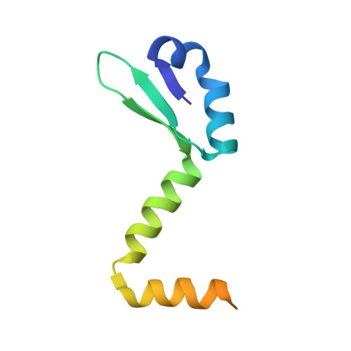
Toxin-antitoxin modules are present on chromosomes of almost all free-living prokaryotes. Some are implicated to act as stress-responsive elements, among their many functional roles. The YefM-YoeB toxin-antitoxin system is present in many bacterial species, where YefM belongs to the Phd family antidote of phage P1, whereas YoeB is a homolog of the RelE toxin of the RelBE system, rather than the Doc system of phage P1. YoeB, a ribonuclease, is believed to be conformationally stable, whereas YefM has been proposed to be a member of intrinsically disordered proteins. The ribonucleolytic activity of YoeB is neutralized by YefM upon formation of the YefM-YoeB complex. We report here the crystal structure of Mycobacterium tuberculosis YefM from two crystal isoforms. Our crystallographic and biophysical studies reveal that YefM is not an intrinsically unfolded protein and instead forms a well-defined structure with significant secondary and tertiary structure conformations. The residues involved in core formation of the folded structure are evolutionarily conserved among many bacterial species, supporting our observation. The C-terminal end of its polypeptide is highly pliable, which adopts different conformations in different monomers. Since at the physiological level YefM controls the activity of YoeB through intricate protein-protein interactions, the conformational heterogeneity in YefM revealed by our structure suggests that these might act a master switch in controlling YoeB activity.
Organizational Affiliation:
Laboratory of Structural Biology, Centre for DNA Fingerprinting and Diagnostics, Nacharam, Hyderabad 500 076, India.