Topology conservation and loop flexibility in quadruplex-drug recognition: crystal structures of inter- and intramolecular telomeric DNA quadruplex-drug complexes
Parkinson, G.N., Cuenca, F., Neidle, S.(2008) J Mol Biol 381: 1145-1156
- PubMed: 18619463
- DOI: https://doi.org/10.1016/j.jmb.2008.06.022
- Primary Citation of Related Structures:
3CCO, 3CDM - PubMed Abstract:
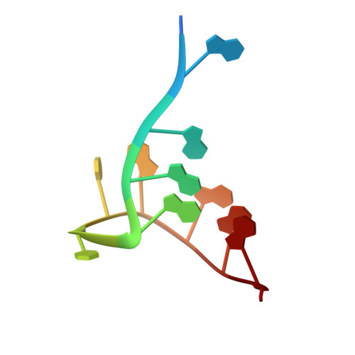
Knowledge of the biologically relevant topology is critical for the design of drugs targeting quadruplex nucleic acids. We report here crystal structures of a G-quadruplex-selective ligand complexed with two human telomeric DNA quadruplexes. The intramolecular quadruplex sequence d[TAGGG(TTAGGG)(3)] and the bimolecular quadruplex sequence d(TAGGGTTAGGGT) were co-crystallized with a tetra-substituted naphthalene diimide quadruplex-binding ligand. The structures were solved and refined to 2.10- and 2.20-A resolution, respectively, revealing that the quadruplex topology in both structures is unchanged by the addition of the ligands, retaining a parallel-stranded arrangement with external double-chain-reversal propeller loops. The parallel topology results in accessible external 5' and 3' planar G-tetrad surfaces for ligand stacking. This also enables significant ligand-induced conformational changes in several TTA propeller loops to take place such that the loops themselves are able to accommodate bound drug molecules without affecting the parallel quadruplex topology, by stacking on the external TTA connecting loop nucleotides. Ligands are bound into the external TTA loop nucleotides and stack onto G-tetrad surfaces. These crystal structures provide a framework for further ligand development of the naphthalene diimide series to enhance selectivity and affinity.
Organizational Affiliation:
The Cancer Research UK Biomolecular Structure Group, The School of Pharmacy, University of London, 29-39 Brunswick Square, London WC1N 1AX, UK.