


Crystal structure of the ternary complex of phospholipase A2 with ajmaline and anisic acid at 3.1 A resolution
Kumar, S., Vikram, G., Singh, N., Sharma, S., Kaur, P., Singh, T.P.To be published.
Experimental Data Snapshot
Entity ID: 1 | |||||
|---|---|---|---|---|---|
| Molecule | Chains | Sequence Length | Organism | Details | Image |
| Phospholipase A2 VRV-PL-VIIIa | 121 | Daboia russelii pulchella | Mutation(s): 0 EC: 3.1.1.4 | 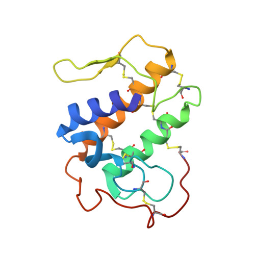 | |
UniProt | |||||
Find proteins for P59071 (Daboia russelii) Explore P59071 Go to UniProtKB: P59071 | |||||
Entity Groups | |||||
| Sequence Clusters | 30% Identity50% Identity70% Identity90% Identity95% Identity100% Identity | ||||
| UniProt Group | P59071 | ||||
Sequence AnnotationsExpand | |||||
| |||||
| Ligands 2 Unique | |||||
|---|---|---|---|---|---|
| ID | Chains | Name / Formula / InChI Key | 2D Diagram | 3D Interactions | |
| AJM Query on AJM | E [auth A], G [auth B], I [auth C], K [auth D] | AJMALINE C18 H22 N2 O2 CFEPCEVMXPTZPJ-OGDRVKPDSA-N |  | ||
| ANN Query on ANN | F [auth A], H [auth B], J [auth C], L [auth D] | 4-METHOXYBENZOIC ACID C8 H8 O3 ZEYHEAKUIGZSGI-UHFFFAOYSA-N |  | ||
| Length ( Å ) | Angle ( ˚ ) |
|---|---|
| a = 108.799 | α = 90 |
| b = 108.799 | β = 90 |
| c = 43.339 | γ = 120 |
| Software Name | Purpose |
|---|---|
| DENZO | data reduction |
| AMoRE | phasing |
| CNS | refinement |
| AUTOMAR | data reduction |
| SCALEPACK | data scaling |
RCSB PDB (citation) is hosted by
RCSB PDB is a member of the

