3CAP

Crystal Structure of Native Opsin: the G Protein-Coupled Receptor Rhodopsin in its Ligand-free State


Experimental Data Snapshot

  • Method: X-RAY DIFFRACTION
  • Resolution: 2.90 Å
  • R-Value Free: 0.266 
  • R-Value Work: 0.229 
  • R-Value Observed: 0.231 

wwPDB Validation   3D Report Full Report


Ligand Structure Quality Assessment 


This is version 2.1 of the entry. See complete history


Literature

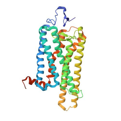
Crystal structure of the ligand-free G-protein-coupled receptor opsin

Park, J.H.Scheerer, P.Hofmann, K.P.Choe, H.-W.Ernst, O.P.

(2008) Nature 454: 183-187

  • DOI: https://doi.org/10.1038/nature07063
  • Primary Citation of Related Structures:  
    3CAP

  • PubMed Abstract: 

    In the G-protein-coupled receptor (GPCR) rhodopsin, the inactivating ligand 11-cis-retinal is bound in the seven-transmembrane helix (TM) bundle and is cis/trans isomerized by light to form active metarhodopsin II. With metarhodopsin II decay, all-trans-retinal is released, and opsin is reloaded with new 11-cis-retinal. Here we present the crystal structure of ligand-free native opsin from bovine retinal rod cells at 2.9 ångström (A) resolution. Compared to rhodopsin, opsin shows prominent structural changes in the conserved E(D)RY and NPxxY(x)(5,6)F regions and in TM5-TM7. At the cytoplasmic side, TM6 is tilted outwards by 6-7 A, whereas the helix structure of TM5 is more elongated and close to TM6. These structural changes, some of which were attributed to an active GPCR state, reorganize the empty retinal-binding pocket to disclose two openings that may serve the entry and exit of retinal. The opsin structure sheds new light on ligand binding to GPCRs and on GPCR activation.


  • Organizational Affiliation

    Institut für Medizinische Physik und Biophysik (CC2), Charité-Universitätsmedizin Berlin, Charitéplatz 1, D-10117 Berlin, Germany.


Macromolecules
Find similar proteins by:  (by identity cutoff)  |  3D Structure
Entity ID: 1
MoleculeChains Sequence LengthOrganismDetailsImage
Rhodopsin
A, B
348Bos taurusMutation(s): 0 
Membrane Entity: Yes 
UniProt
Find proteins for P02699 (Bos taurus)
Explore P02699 
Go to UniProtKB:  P02699
Entity Groups  
Sequence Clusters30% Identity50% Identity70% Identity90% Identity95% Identity100% Identity
UniProt GroupP02699
Sequence Annotations
Expand
  • Reference Sequence
Oligosaccharides

Help

Entity ID: 2
MoleculeChains Length2D Diagram Glycosylation3D Interactions
beta-D-mannopyranose-(1-3)-beta-D-mannopyranose-(1-4)-2-acetamido-2-deoxy-beta-D-glucopyranose-(1-4)-2-acetamido-2-deoxy-beta-D-glucopyranose
C, E
4N-Glycosylation
Glycosylation Resources
GlyTouCan:  G31886NL
GlyCosmos:  G31886NL
GlyGen:  G31886NL
Entity ID: 3
MoleculeChains Length2D Diagram Glycosylation3D Interactions
2-acetamido-2-deoxy-beta-D-glucopyranose-(1-4)-2-acetamido-2-deoxy-beta-D-glucopyranose
D, F
2N-Glycosylation
Glycosylation Resources
GlyTouCan:  G42666HT
GlyCosmos:  G42666HT
GlyGen:  G42666HT
Experimental Data & Validation

Experimental Data

  • Method: X-RAY DIFFRACTION
  • Resolution: 2.90 Å
  • R-Value Free: 0.266 
  • R-Value Work: 0.229 
  • R-Value Observed: 0.231 
  • Space Group: H 3
Unit Cell:
Length ( Å )Angle ( ˚ )
a = 242.917α = 90
b = 242.918β = 90
c = 110.42γ = 120
Software Package:
Software NamePurpose
MAR345dtbdata collection
CNSrefinement
REFMACrefinement
HKL-2000data reduction
HKL-2000data scaling
PHASERphasing

Structure Validation

View Full Validation Report


Ligand Structure Quality Assessment 


Entry History 

Deposition Data

Revision History  (Full details and data files)

  • Version 1.0: 2008-06-24
    Type: Initial release
  • Version 1.1: 2011-07-13
    Changes: Non-polymer description, Version format compliance
  • Version 2.0: 2020-07-29
    Type: Remediation
    Reason: Carbohydrate remediation
    Changes: Advisory, Atomic model, Data collection, Derived calculations, Structure summary
  • Version 2.1: 2023-11-01
    Changes: Data collection, Database references, Refinement description, Structure summary