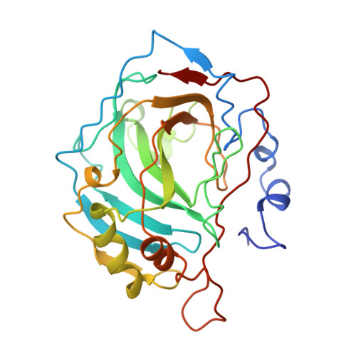
Crystallographic studies of inhibitor binding sites in human carbonic anhydrase II: a pentacoordinated binding of the SCN- ion to the zinc at high pH.
Eriksson, A.E., Kylsten, P.M., Jones, T.A., Liljas, A.(1988) Proteins 4: 283-293
- PubMed: 3151020
- DOI: https://doi.org/10.1002/prot.340040407
- Primary Citation of Related Structures:
2CA2, 3CA2 - PubMed Abstract:
The binding of four inhibitors--mercuric ion, 3-acetoxymercuri-4-aminobenzenesulfonamide (AMS), acetazolamide (Diamox), and thiocyanate ion--to human carbonic anhydrase II (HCA II) has been studied with X-ray crystallography. The binding of mercury to HCA II at pH 7.0 has been investigated at 3.1 A resolution. Mercuric ions are observed at both nitrogens in the His-64 ring. One of these sites is pointing toward the zinc ion. The only other binding site for mercury is at Cys-206. The binding of the two sulfonamide inhibitors AMS and Diamox, has been reinvestigated at 2.0 and 3.0 A, respectively. Only the nitrogen of the sulfonamide group binds to the zinc ion replacing the hydroxyl ion. The sulfonamide oxygen closest to the zinc ion is 3.1 A away. Thus the tetrahedral geometry of the zinc is retained, refuting earlier models of a pentacoordinated zinc. The structure of the thiocyanate complex has been investigated at pH 8.5 and the structure has been refined at 1.9 A resolution using the least-squares refinement program PROLSQ. The crystallographic R factor is 17.6%. The zinc ion is pentacoordinated with the anion as well as a water molecule bound in addition to the three histidine residues. The nitrogen atom of the SCN- ion is 1.9 A from the zinc ion but shifted 1.3 A with respect to the hydroxyl ion in the native structure and at van der Waals' distance from the O gamma l atom of Thr-199. This is due to the inability of the O gamma l atom of Thr-199 to serve as a hydrogen bond donor, thus repelling the nonprotonated nitrogen. The SCN- molecule reaches into the deep end of the active site cavity where the sulfur atom has displaced the so-called "deep" water molecule of the native enzyme. The zinc-bound water molecule is 2.2 A from the zinc ion and 2.4 A from the SCN- nitrogen. In addition, this water is hydrogen bonded to the O gamma l atom of Thr-199 and to another water molecule. We have observed that solvent and inhibitor molecules have three possible binding sites on the zinc ion and their significance for the catalysis and inhibition of HCA II will be discussed. All available crystallographic data are consistent with a proposed catalytic mechanism in which both the OH moiety and one oxygen of the substrate HCO3- ion are ligated to the zinc ion.
Organizational Affiliation:
Department of Molecular Biology, Biomedical Center, Uppsala, Sweden.