


The crystal structure of a putative membrane protein from Corynebacterium diphtheriae.
Zhang, R., Hatzos, C., Clancy, S., Joachimiak, A.To be published.
Experimental Data Snapshot
wwPDB Validation 3D Report Full Report

Entity ID: 1 | |||||
|---|---|---|---|---|---|
| Molecule | Chains | Sequence Length | Organism | Details | Image |
| Putative membrane protein | 141 | Corynebacterium diphtheriae NCTC 13129 | Mutation(s): 0 Gene Names: GI:38199630, DIP0771 | 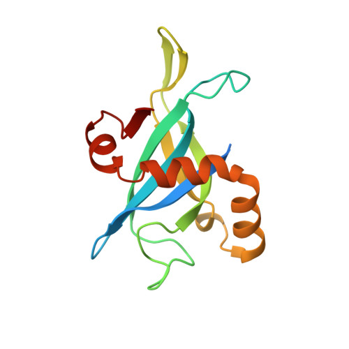 | |
UniProt | |||||
Find proteins for Q6NIK4 (Corynebacterium diphtheriae (strain ATCC 700971 / NCTC 13129 / Biotype gravis)) Explore Q6NIK4 Go to UniProtKB: Q6NIK4 | |||||
Entity Groups | |||||
| Sequence Clusters | 30% Identity50% Identity70% Identity90% Identity95% Identity100% Identity | ||||
| UniProt Group | Q6NIK4 | ||||
Sequence AnnotationsExpand | |||||
| |||||
| Length ( Å ) | Angle ( ˚ ) |
|---|---|
| a = 50.374 | α = 90 |
| b = 99.837 | β = 90 |
| c = 124.621 | γ = 90 |
| Software Name | Purpose |
|---|---|
| REFMAC | refinement |
| SBC-Collect | data collection |
| HKL-3000 | data reduction |
| HKL-3000 | data scaling |
| HKL-3000 | phasing |
RCSB PDB (citation) is hosted by
RCSB PDB is a member of the

