Computational redesign of the SHV-1 beta-lactamase/beta-lactamase inhibitor protein interface.
Reynolds, K.A., Hanes, M.S., Thomson, J.M., Antczak, A.J., Berger, J.M., Bonomo, R.A., Kirsch, J.F., Handel, T.M.(2008) J Mol Biol 382: 1265-1275
- PubMed: 18775544
- DOI: https://doi.org/10.1016/j.jmb.2008.05.051
- Primary Citation of Related Structures:
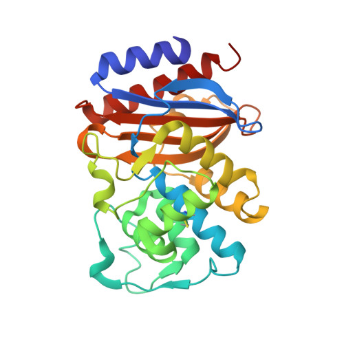
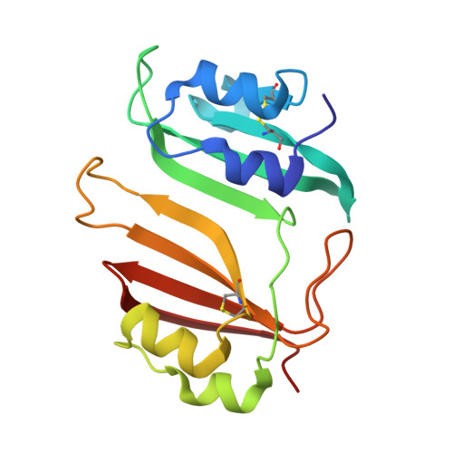
3C4O, 3C4P - PubMed Abstract:
Beta-lactamases are enzymes that catalyze the hydrolysis of beta-lactam antibiotics. beta-lactamase/beta-lactamase inhibitor protein (BLIP) complexes are emerging as a well characterized experimental model system for studying protein-protein interactions. BLIP is a 165 amino acid protein that inhibits several class A beta-lactamases with a wide range of affinities: picomolar affinity for K1; nanomolar affinity for TEM-1, SME-1, and BlaI; but only micromolar affinity for SHV-1 beta-lactamase. The large differences in affinity coupled with the availability of extensive mutagenesis data and high-resolution crystal structures for the TEM-1/BLIP and SHV-1/BLIP complexes make them attractive systems for the further development of computational design methodology. We used EGAD, a physics-based computational design program, to redesign BLIP in an attempt to increase affinity for SHV-1. Characterization of several of designs and point mutants revealed that in all cases, the mutations stabilize the interface by 10- to 1000-fold relative to wild type BLIP. The calculated changes in binding affinity for the mutants were within a mean absolute error of 0.87 kcal/mol from the experimental values, and comparison of the calculated and experimental values for a set of 30 SHV-1/BLIP complexes yielded a correlation coefficient of 0.77. Structures of the two complexes with the highest affinity, SHV-1/BLIP (E73M) and SHV-1/BLIP (E73M, S130K, S146M), are presented at 1.7 A resolution. While the predicted structures have much in common with the experimentally determined structures, they do not coincide perfectly; in particular a salt bridge between SHV-1 D104 and BLIP K74 is observed in the experimental structures, but not in the predicted design conformations. This discrepancy highlights the difficulty of modeling salt bridge interactions with a protein design algorithm that approximates side chains as discrete rotamers. Nevertheless, while local structural features of the interface were sometimes miscalculated, EGAD is globally successful in designing complexes with increased affinity.
Organizational Affiliation:
Biophysics Graduate Group, University of California, Berkeley, Berkeley, CA 94720, USA.