3C2U

Structure of the two subsite D-xylosidase from Selenomonas ruminantium in complex with 1,3-bis[tris(hydroxymethyl)methylamino]propane


Experimental Data Snapshot

  • Method: X-RAY DIFFRACTION
  • Resolution: 1.30 Å
  • R-Value Free: 0.163 
  • R-Value Work: 0.134 
  • R-Value Observed: 0.136 

wwPDB Validation   3D Report Full Report


This is version 1.3 of the entry. See complete history


Literature

Structure of the two-subsite beta-d-xylosidase from Selenomonas ruminantium in complex with 1,3-bis[tris(hydroxymethyl)methylamino]propane.

Brunzelle, J.S.Jordan, D.B.McCaslin, D.R.Olczak, A.Wawrzak, Z.

(2008) Arch Biochem Biophys 474: 157-166

  • DOI: https://doi.org/10.1016/j.abb.2008.03.007
  • Primary Citation of Related Structures:  
    3C2U

  • PubMed Abstract: 

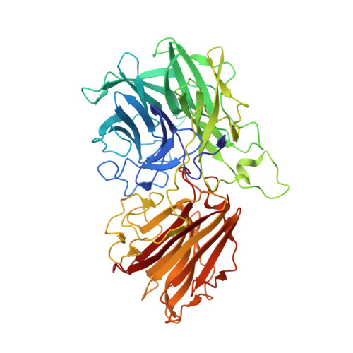
    The three-dimensional structure of the catalytically efficient beta-xylosidase from Selenomonas ruminantium in complex with competitive inhibitor 1,3-bis[tris(hydroxymethyl)methylamino]propane (BTP) was determined by using X-ray crystallography (1.3A resolution). Most H bonds between inhibitor and protein occur within subsite -1, including one between the carboxyl group of E186 and an N group of BTP. The other N of BTP occupies subsite +1 near K99. E186 (pK(a) 7.2) serves as catalytic acid. The pH (6-10) profile for 1/K(i)((BTP)) is bell-shaped with pK(a)'s 6.8 and 7.8 on the acidic limb assigned to E186 and inhibitor groups and 9.9 on the basic limb assigned to inhibitor. Mutation K99A eliminates pK(a) 7.8, strongly suggesting that the BTP monocation binds to the dianionic enzyme D14(-)E186(-). A sedimentation equilibrium experiment estimates a K(d) ([dimer](2)/[tetramer]) of 7 x 10(-9)M. Similar k(cat) and k(cat)/K(m) values were determined when the tetramer/dimer ratio changes from 0.0028 to 26 suggesting that dimers and tetramers are equally active forms.


  • Organizational Affiliation

    Northwestern University Center for Synchrotron Research, Life Sciences Collaborative Access Team, Department of Molecular Pharmacology and Biological Chemistry, 9700 South Cass Avenue, Argonne, IL 60439, USA.


Macromolecules
Find similar proteins by:  (by identity cutoff)  |  3D Structure
Entity ID: 1
MoleculeChains Sequence LengthOrganismDetailsImage
Xylosidase/arabinosidase
A, B, C, D
538Selenomonas ruminantiumMutation(s): 0 
UniProt
Find proteins for O52575 (Selenomonas ruminantium)
Explore O52575 
Go to UniProtKB:  O52575
Entity Groups  
Sequence Clusters30% Identity50% Identity70% Identity90% Identity95% Identity100% Identity
UniProt GroupO52575
Sequence Annotations
Expand
  • Reference Sequence
Small Molecules
Binding Affinity Annotations 
IDSourceBinding Affinity
BTP PDBBind:  3C2U Ki: 1.25e+6 (nM) from 1 assay(s)
Experimental Data & Validation

Experimental Data

  • Method: X-RAY DIFFRACTION
  • Resolution: 1.30 Å
  • R-Value Free: 0.163 
  • R-Value Work: 0.134 
  • R-Value Observed: 0.136 
  • Space Group: P 1
Unit Cell:
Length ( Å )Angle ( ˚ )
a = 77.579α = 67.84
b = 84.401β = 81.31
c = 94.039γ = 75.08
Software Package:
Software NamePurpose
REFMACrefinement
MAR345data collection
XDSdata reduction
XDSdata scaling
XSCALEdata scaling
SnBphasing

Structure Validation

View Full Validation Report


Entry History 

Deposition Data

Revision History  (Full details and data files)

  • Version 1.0: 2008-04-22
    Type: Initial release
  • Version 1.1: 2011-07-13
    Changes: Version format compliance
  • Version 1.2: 2017-10-25
    Changes: Refinement description
  • Version 1.3: 2024-02-21
    Changes: Data collection, Database references, Derived calculations