3BUJ

Crystal Structure of CalO2


Experimental Data Snapshot

  • Method: X-RAY DIFFRACTION
  • Resolution: 2.47 Å
  • R-Value Free: 0.257 
  • R-Value Work: 0.198 
  • R-Value Observed: 0.201 

wwPDB Validation   3D Report Full Report


Ligand Structure Quality Assessment 


This is version 1.4 of the entry. See complete history


Literature

Structural characterization of CalO2: a putative orsellinic acid P450 oxidase in the calicheamicin biosynthetic pathway.

McCoy, J.G.Johnson, H.D.Singh, S.Bingman, C.A.Lei, I.K.Thorson, J.S.Phillips Jr., G.N.

(2009) Proteins 74: 50-60

  • DOI: https://doi.org/10.1002/prot.22131
  • Primary Citation of Related Structures:  
    3BUJ

  • PubMed Abstract: 

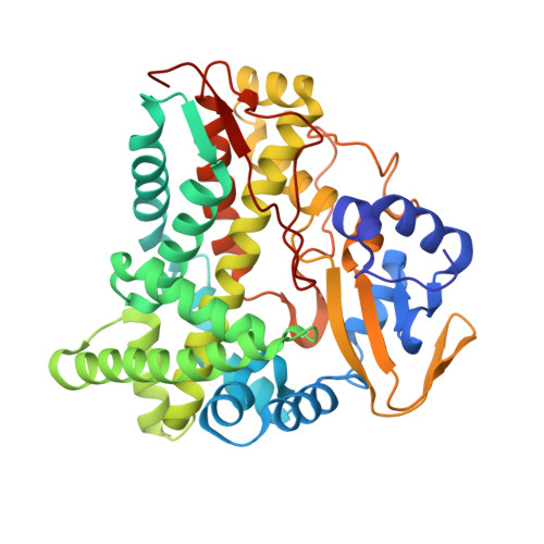
    Although bacterial iterative Type I polyketide synthases are now known to participate in the biosynthesis of a small set of diverse natural products, the subsequent downstream modification of the resulting polyketide products remains poorly understood. Toward this goal, we report the X-ray structure determination at 2.5 A resolution and preliminary characterization of the putative orsellenic acid P450 oxidase (CalO2) involved in calicheamicin biosynthesis. These studies represent the first crystal structure for a P450 involved in modifying a bacterial iterative Type I polyketide product and suggest the CalO2-catalyzed step may occur after CalO3-catalyzed iodination and may also require a coenzyme A- (CoA) or acyl carrier protein- (ACP) bound substrate. Docking studies also reveal a putative docking site within CalO2 for the CLM orsellinic acid synthase (CalO5) ACP domain which involves a well-ordered helix along the CalO2 active site cavity that is unique compared with other P450 structures.


  • Organizational Affiliation

    Department of Biochemistry, University of Wisconsin-Madison, Madison, Wisconsin 53706-1544, USA.


Macromolecules
Find similar proteins by:  (by identity cutoff)  |  3D Structure
Entity ID: 1
MoleculeChains Sequence LengthOrganismDetailsImage
CalO2397Micromonospora echinosporaMutation(s): 0 
Gene Names: calO2
UniProt
Find proteins for Q8KND6 (Micromonospora echinospora)
Explore Q8KND6 
Go to UniProtKB:  Q8KND6
Entity Groups  
Sequence Clusters30% Identity50% Identity70% Identity90% Identity95% Identity100% Identity
UniProt GroupQ8KND6
Sequence Annotations
Expand
  • Reference Sequence
Small Molecules
Ligands 1 Unique
IDChains Name / Formula / InChI Key2D Diagram3D Interactions
HEM
Query on HEM

Download Ideal Coordinates CCD File 
B [auth A]PROTOPORPHYRIN IX CONTAINING FE
C34 H32 Fe N4 O4
KABFMIBPWCXCRK-RGGAHWMASA-L
Experimental Data & Validation

Experimental Data

  • Method: X-RAY DIFFRACTION
  • Resolution: 2.47 Å
  • R-Value Free: 0.257 
  • R-Value Work: 0.198 
  • R-Value Observed: 0.201 
  • Space Group: P 41 21 2
Unit Cell:
Length ( Å )Angle ( ˚ )
a = 71.202α = 90
b = 71.202β = 90
c = 204.451γ = 90
Software Package:
Software NamePurpose
DENZOdata reduction
SCALEPACKdata scaling
MOLREPphasing
DMphasing
REFMACrefinement
PDB_EXTRACTdata extraction

Structure Validation

View Full Validation Report


Ligand Structure Quality Assessment 


Entry History 

Deposition Data

Revision History  (Full details and data files)

  • Version 1.0: 2008-04-29
    Type: Initial release
  • Version 1.1: 2011-07-13
    Changes: Advisory, Version format compliance
  • Version 1.2: 2013-01-30
    Changes: Database references, Structure summary
  • Version 1.3: 2017-10-25
    Changes: Refinement description
  • Version 1.4: 2023-08-30
    Changes: Data collection, Database references, Derived calculations, Refinement description