3BNR

Crystal Structure of the Homo sapiens Mitochondrial Ribosomal Decoding Site in the presence of nonspecifically bound paromomycin (A1555G mutant, Br-derivative)

  • Classification: RNA
  • Mutation(s): No 

  • Deposited: 2007-12-14 Released: 2008-06-24 
  • Deposition Author(s): Kondo, J., Westhof, E.

Experimental Data Snapshot

  • Method: X-RAY DIFFRACTION
  • Resolution: 2.10 Å
  • R-Value Free: 0.245 
  • R-Value Work: 0.215 

wwPDB Validation   3D Report Full Report


Ligand Structure Quality Assessment 


This is version 1.2 of the entry. See complete history


Literature

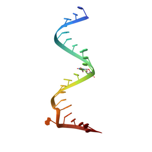
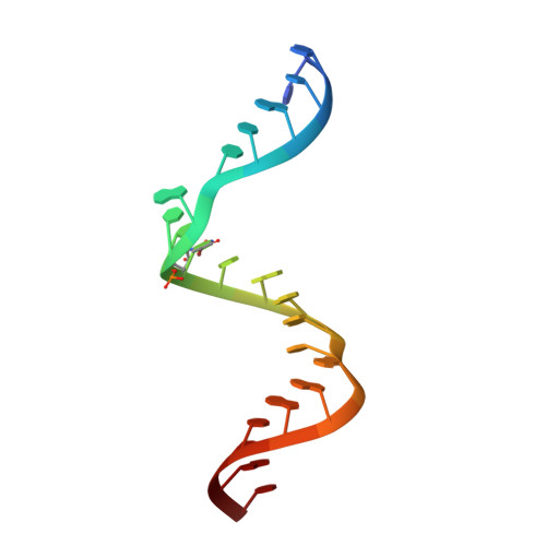
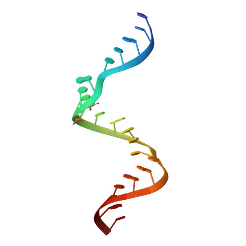
The bacterial and mitochondrial ribosomal A-site molecular switches possess different conformational substates

Kondo, J.Westhof, E.

(2008) Nucleic Acids Res 36: 2654-2666

  • DOI: https://doi.org/10.1093/nar/gkn112
  • Primary Citation of Related Structures:  
    3BNL, 3BNN, 3BNO, 3BNP, 3BNQ, 3BNR, 3BNS, 3BNT

  • PubMed Abstract: 

    The A site of the small ribosomal subunit participates in the fidelity of decoding by switching between two states, a resting 'off' state and an active decoding 'on' state. Eight crystal structures of RNA duplexes containing two minimal decoding A sites of the Homo sapiens mitochondrial wild-type, the A1555G mutant or bacteria have been solved. The resting 'off' state of the mitochondrial wild-type A site is surprisingly different from that of the bacterial A site. The mitochondrial A1555G mutant has two types of the 'off' states; one is similar to the mitochondrial wild-type 'off' state and the other is similar to the bacterial 'off' state. Our present results indicate that the dynamics of the A site in bacteria and mitochondria are different, a property probably related to the small number of tRNAs used for decoding in mitochondria. Based on these structures, we propose a hypothesis for the molecular mechanism of non-syndromic hearing loss due to the mitochondrial A1555G mutation.


  • Organizational Affiliation

    Architecture et Réactivité de l'ARN, Université Louis Pasteur, Institut de Biologie Moléculaire et Cellulaire, CNRS, 15 rue René Descartes, 67084 Strasbourg, France.


Macromolecules

Find similar nucleic acids by:  Sequence   |   3D Structure  

Entity ID: 1
MoleculeChains LengthOrganismImage
A site of human mitochondrial ribosome, chain one
A, C
23N/A
Sequence Annotations
Expand
  • Reference Sequence

Find similar nucleic acids by:  Sequence   |   3D Structure  

Entity ID: 2
MoleculeChains LengthOrganismImage
A site of human mitochondrial ribosome, chain two22N/A
Sequence Annotations
Expand
  • Reference Sequence

Find similar nucleic acids by:  Sequence   |   3D Structure  

Entity ID: 3
MoleculeChains LengthOrganismImage
A site of human mitochondrial ribosome, chain three21N/A
Sequence Annotations
Expand
  • Reference Sequence
Experimental Data & Validation

Experimental Data

  • Method: X-RAY DIFFRACTION
  • Resolution: 2.10 Å
  • R-Value Free: 0.245 
  • R-Value Work: 0.215 
  • Space Group: C 1 2 1
Unit Cell:
Length ( Å )Angle ( ˚ )
a = 71.189α = 90
b = 77.38β = 115.12
c = 56.322γ = 90
Software Package:
Software NamePurpose
SCALAdata scaling
AMoREphasing
CNSrefinement
PDB_EXTRACTdata extraction

Structure Validation

View Full Validation Report


Ligand Structure Quality Assessment 


Entry History 

Deposition Data

Revision History  (Full details and data files)

  • Version 1.0: 2008-06-24
    Type: Initial release
  • Version 1.1: 2011-07-13
    Changes: Version format compliance
  • Version 1.2: 2024-02-21
    Changes: Data collection, Database references, Derived calculations, Structure summary