3BFU

Structure of the ligand-binding core of GluR2 in complex with the agonist (R)-TDPA at 1.95 A resolution


Experimental Data Snapshot

  • Method: X-RAY DIFFRACTION
  • Resolution: 1.95 Å
  • R-Value Free: 0.233 
  • R-Value Work: 0.200 

wwPDB Validation   3D Report Full Report


Ligand Structure Quality Assessment 


This is version 1.4 of the entry. See complete history


Literature

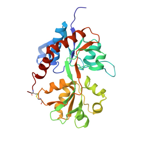
Structures of the ligand-binding core of iGluR2 in complex with the agonists (R)- and (S)-2-amino-3-(4-hydroxy-1,2,5-thiadiazol-3-yl)propionic acid explain their unusual equipotency.

Beich-Frandsen, M.Pickering, D.S.Mirza, O.Johansen, T.N.Greenwood, J.Vestergaard, B.Schousboe, A.Gajhede, M.Liljefors, T.Kastrup, J.S.

(2008) J Med Chem 51: 1459-1463

  • DOI: https://doi.org/10.1021/jm701126w
  • Primary Citation of Related Structures:  
    3BFT, 3BFU

  • PubMed Abstract: 

    AMPA-type ionotropic glutamate receptors generally display high stereoselectivity in agonist binding. However, the stereoisomers of 2-amino-3-(4-hydroxy-1,2,5-thiadiazol-3-yl)propionic acid (TDPA) have similar enantiopharmacology. To understand this observation, we have determined the X-ray structures of ( R)-TDPA and ( S)-TDPA in complex with the ligand-binding core of iGluR2 and investigated the binding pharmacology at AMPA and kainate receptors. Both enantiomers induce full domain closure in iGluR2 but adopt different conformations when binding to the receptor, which may explain the similar enantiopharmacology.


  • Organizational Affiliation

    Department of Medicinal Chemistry, University of Copenhagen, Copenhagen, Denmark.


Macromolecules
Find similar proteins by:  (by identity cutoff)  |  3D Structure
Entity ID: 1
MoleculeChains Sequence LengthOrganismDetailsImage
Glutamate receptor 2
A, B, C, D
263Rattus norvegicusMutation(s): 0 
Gene Names: Gria2Glur2
UniProt
Find proteins for P19491 (Rattus norvegicus)
Explore P19491 
Go to UniProtKB:  P19491
Entity Groups  
Sequence Clusters30% Identity50% Identity70% Identity90% Identity95% Identity100% Identity
UniProt GroupP19491
Sequence Annotations
Expand
  • Reference Sequence
Small Molecules
Binding Affinity Annotations 
IDSourceBinding Affinity
R2P PDBBind:  3BFU Ki: 531 (nM) from 1 assay(s)
Binding MOAD:  3BFU Ki: 531 (nM) from 1 assay(s)
Experimental Data & Validation

Experimental Data

  • Method: X-RAY DIFFRACTION
  • Resolution: 1.95 Å
  • R-Value Free: 0.233 
  • R-Value Work: 0.200 
  • Space Group: P 1 2 1
Unit Cell:
Length ( Å )Angle ( ˚ )
a = 104.75α = 90
b = 47.52β = 113.54
c = 124.33γ = 90
Software Package:
Software NamePurpose
XSCALEdata scaling
MOLREPphasing
CNSrefinement
PDB_EXTRACTdata extraction
xia2data scaling
XDSdata scaling

Structure Validation

View Full Validation Report


Ligand Structure Quality Assessment 


Entry History 

Deposition Data

Revision History  (Full details and data files)

  • Version 1.0: 2008-10-14
    Type: Initial release
  • Version 1.1: 2011-07-13
    Changes: Version format compliance
  • Version 1.2: 2017-08-23
    Changes: Refinement description, Source and taxonomy
  • Version 1.3: 2017-10-25
    Changes: Refinement description
  • Version 1.4: 2023-11-01
    Changes: Data collection, Database references, Derived calculations, Refinement description