3BE7

Crystal structure of Zn-dependent arginine carboxypeptidase


Experimental Data Snapshot

  • Method: X-RAY DIFFRACTION
  • Resolution: 2.30 Å
  • R-Value Free: 0.280 
  • R-Value Work: 0.226 
  • R-Value Observed: 0.228 

wwPDB Validation   3D Report Full Report


Ligand Structure Quality Assessment 


This is version 1.4 of the entry. See complete history


Literature

Functional identification of incorrectly annotated prolidases from the amidohydrolase superfamily of enzymes.

Xiang, D.F.Patskovsky, Y.Xu, C.Meyer, A.J.Sauder, J.M.Burley, S.K.Almo, S.C.Raushel, F.M.

(2009) Biochemistry 48: 3730-3742

  • DOI: https://doi.org/10.1021/bi900111q
  • Primary Citation of Related Structures:  
    3BE7, 3DUG

  • PubMed Abstract: 

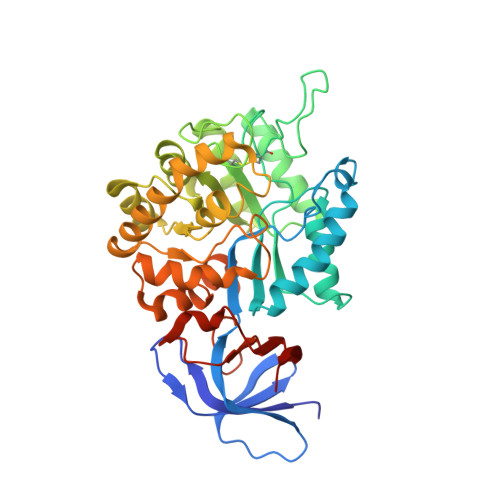
    The substrate profiles for two proteins from Caulobacter crescentus CB15 (Cc2672 and Cc3125) and one protein (Sgx9359b) derived from a DNA sequence ( gi|44368820 ) isolated from the Sargasso Sea were determined using combinatorial libraries of dipeptides and N-acyl derivatives of amino acids. These proteins are members of the amidohydrolase superfamily and are currently misannotated in NCBI as catalyzing the hydrolysis of l-Xaa-l-Pro dipeptides. Cc2672 was shown to catalyze the hydrolysis of l-Xaa-l-Arg/Lys dipeptides and the N-acetyl and N-formyl derivatives of lysine and arginine. This enzyme will also hydrolyze longer peptides that terminate in either lysine or arginine. The N-methyl phosphonate derivative of l-lysine was a potent competitive inhibitor of Cc2672 with a K(i) value of 120 nM. Cc3125 was shown to catalyze the hydrolysis of l-Xaa-l-Arg/Lys dipeptides but will not hydrolyze tripeptides or the N-formyl and N-acetyl derivatives of lysine or arginine. The substrate profile for Sgx9359b is similar to that of Cc2672 except that compounds with a C-terminal lysine are not recognized as substrates. The X-ray structure of Sgx9359b was determined to a resolution of 2.3 A. The protein folds as a (beta/alpha)(8)-barrel and self-associates to form a homooctamer. The active site is composed of a binuclear metal center similar to that found in phosphotriesterase and dihydroorotase. In one crystal form, arginine was bound adventitiously to the eight active sites within the octamer. The orientation of the arginine in the active site identified the structural determinants for recognition of the alpha-carboxylate and the positively charged side chains of arginine-containing substrates. This information was used to identify 18 other bacterial sequences that possess identical or similar substrate profiles.


  • Organizational Affiliation

    Department of Chemistry, Texas A&M University, P.O. Box 30012, College Station, Texas 77842-3012, USA.


Macromolecules
Find similar proteins by:  (by identity cutoff)  |  3D Structure
Entity ID: 1
MoleculeChains Sequence LengthOrganismDetailsImage
Zn-dependent arginine carboxypeptidase
A, B, C, D, E
A, B, C, D, E, F, G, H
408unidentifiedMutation(s): 0 
Gene Names: IBEA_CTG=2118574
Entity Groups  
Sequence Clusters30% Identity50% Identity70% Identity90% Identity95% Identity100% Identity
Sequence Annotations
Expand
  • Reference Sequence
Small Molecules
Ligands 3 Unique
IDChains Name / Formula / InChI Key2D Diagram3D Interactions
ARG
Query on ARG

Download Ideal Coordinates CCD File 
AA [auth G]
DA [auth H]
I [auth A]
L [auth B]
N [auth C]
AA [auth G],
DA [auth H],
I [auth A],
L [auth B],
N [auth C],
P [auth D],
S [auth E],
W [auth F]
ARGININE
C6 H15 N4 O2
ODKSFYDXXFIFQN-BYPYZUCNSA-O
GOL
Query on GOL

Download Ideal Coordinates CCD File 
BA [auth G]
EA [auth H]
FA [auth H]
GA [auth H]
HA [auth H]
BA [auth G],
EA [auth H],
FA [auth H],
GA [auth H],
HA [auth H],
J [auth A],
K [auth A],
M [auth B],
O [auth C],
Q [auth D],
R [auth D],
T [auth E],
U [auth E],
X [auth F],
Y [auth F],
Z [auth F]
GLYCEROL
C3 H8 O3
PEDCQBHIVMGVHV-UHFFFAOYSA-N
MG
Query on MG

Download Ideal Coordinates CCD File 
CA [auth H],
V [auth F]
MAGNESIUM ION
Mg
JLVVSXFLKOJNIY-UHFFFAOYSA-N
Experimental Data & Validation

Experimental Data

  • Method: X-RAY DIFFRACTION
  • Resolution: 2.30 Å
  • R-Value Free: 0.280 
  • R-Value Work: 0.226 
  • R-Value Observed: 0.228 
  • Space Group: P 21 21 21
Unit Cell:
Length ( Å )Angle ( ˚ )
a = 113.42α = 90
b = 146.583β = 90
c = 255.963γ = 90
Software Package:
Software NamePurpose
MOLREPphasing
REFMACrefinement
HKL-2000data reduction
HKL-2000data scaling

Structure Validation

View Full Validation Report


Ligand Structure Quality Assessment 


Entry History 

Revision History  (Full details and data files)

  • Version 1.0: 2007-11-27
    Type: Initial release
  • Version 1.1: 2011-07-13
    Changes: Non-polymer description, Version format compliance
  • Version 1.2: 2018-11-14
    Changes: Data collection, Structure summary
  • Version 1.3: 2021-02-03
    Changes: Database references, Derived calculations, Structure summary
  • Version 1.4: 2023-08-30
    Changes: Data collection, Database references, Refinement description