3B4N

Crystal Structure Analysis of Pectate Lyase PelI from Erwinia chrysanthemi


Experimental Data Snapshot

  • Method: X-RAY DIFFRACTION
  • Resolution: 1.45 Å
  • R-Value Free: 0.198 
  • R-Value Work: 0.175 
  • R-Value Observed: 0.178 

wwPDB Validation   3D Report Full Report


This is version 1.2 of the entry. See complete history


Literature

The Crystal Structure of Pectate Lyase PelI from Soft Rot Pathogen Erwinia chrysanthemi in Complex with Its Substrate.

Creze, C.Castang, S.Derivery, E.Haser, R.Hugouvieux-Cotte-Pattat, N.Shevchik, V.E.Gouet, P.

(2008) J Biol Chem 283: 18260-18268

  • DOI: https://doi.org/10.1074/jbc.M709931200
  • Primary Citation of Related Structures:  
    3B4N, 3B90

  • PubMed Abstract: 

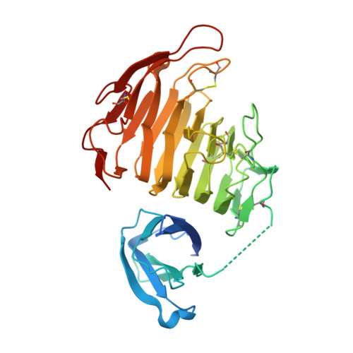
    The crystallographic structure of the family 3 polysaccharide lyase (PL-3) PelI from Erwinia chrysanthemi has been solved to 1.45 A resolution. It consists of an N-terminal domain harboring a fibronectin type III fold linked to a catalytic domain displaying a parallel beta-helix topology. The N-terminal domain is located away from the active site and is not involved in the catalytic process. After secretion in planta, the two domains are separated by E. chrysanthemi proteases. This event turns on the hypersensitive response of the host. The structure of the single catalytic domain determined to 2.1 A resolution shows that the domain separation unveils a "Velcro"-like motif of asparagines, which might be recognized by a plant receptor. The structure of PelI in complex with its substrate, a tetragalacturonate, has been solved to 2.3 A resolution. The sugar binds from subsites -2 to +2 in one monomer of the asymmetric unit, although it lies on subsites -1 to +3 in the other. These two "Michaelis complexes" have never been observed simultaneously before and are consistent with the dual mode of bond cleavage in this substrate. The bound sugar adopts a mixed 2(1) and 3(1) helical conformation similar to that reported in inactive mutants from families PL-1 and PL-10. However, our study suggests that the catalytic base in PelI is not a conventional arginine but a lysine as proposed in family PL-9.


  • Organizational Affiliation

    Laboratoire de BioCristallographie, Institut de Biologie et Chimie des Protéines, CNRS et Université de Lyon, UMR 5086, IFR 128 BioSciences Gerland-Lyon Sud, Lyon, France.


Macromolecules
Find similar proteins by:  (by identity cutoff)  |  3D Structure
Entity ID: 1
MoleculeChains Sequence LengthOrganismDetailsImage
Endo-pectate lyase
A, B
344Dickeya chrysanthemiMutation(s): 0 
Gene Names: pelI
EC: 4.2.2.2
UniProt
Find proteins for O50325 (Dickeya chrysanthemi)
Explore O50325 
Go to UniProtKB:  O50325
Entity Groups  
Sequence Clusters30% Identity50% Identity70% Identity90% Identity95% Identity100% Identity
UniProt GroupO50325
Sequence Annotations
Expand
  • Reference Sequence
Small Molecules
Ligands 4 Unique
IDChains Name / Formula / InChI Key2D Diagram3D Interactions
SO4
Query on SO4

Download Ideal Coordinates CCD File 
N [auth B]SULFATE ION
O4 S
QAOWNCQODCNURD-UHFFFAOYSA-L
ZN
Query on ZN

Download Ideal Coordinates CCD File 
D [auth A]
E [auth A]
F [auth A]
K [auth B]
L [auth B]
D [auth A],
E [auth A],
F [auth A],
K [auth B],
L [auth B],
M [auth B]
ZINC ION
Zn
PTFCDOFLOPIGGS-UHFFFAOYSA-N
EDO
Query on EDO

Download Ideal Coordinates CCD File 
G [auth A]
H [auth A]
I [auth A]
O [auth B]
P [auth B]
G [auth A],
H [auth A],
I [auth A],
O [auth B],
P [auth B],
Q [auth B],
R [auth B]
1,2-ETHANEDIOL
C2 H6 O2
LYCAIKOWRPUZTN-UHFFFAOYSA-N
CA
Query on CA

Download Ideal Coordinates CCD File 
C [auth A],
J [auth B]
CALCIUM ION
Ca
BHPQYMZQTOCNFJ-UHFFFAOYSA-N
Experimental Data & Validation

Experimental Data

  • Method: X-RAY DIFFRACTION
  • Resolution: 1.45 Å
  • R-Value Free: 0.198 
  • R-Value Work: 0.175 
  • R-Value Observed: 0.178 
  • Space Group: P 1 21 1
Unit Cell:
Length ( Å )Angle ( ˚ )
a = 61.6α = 90
b = 70.7β = 112.8
c = 73.4γ = 90
Software Package:
Software NamePurpose
CNSrefinement
DNAdata collection
XDSdata reduction
XSCALEdata scaling
SOLVEphasing

Structure Validation

View Full Validation Report


Entry History 

Deposition Data

Revision History  (Full details and data files)

  • Version 1.0: 2008-04-29
    Type: Initial release
  • Version 1.1: 2011-07-13
    Changes: Version format compliance
  • Version 1.2: 2024-04-03
    Changes: Data collection, Database references, Derived calculations, Refinement description