Conformational dynamics of wild-type Lys48-linked diubiquitin in solution
Hirano, T., Serve, O., Yagi-Utsumi, M., Takemoto, E., Hiromoto, T., Satoh, T., Mizushima, T., Kato, K.(2011) J Biol Chem 286: 37496-37502
- PubMed: 21900242
- DOI: https://doi.org/10.1074/jbc.M111.256354
- Primary Citation of Related Structures:
3AUL - PubMed Abstract:
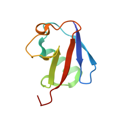
Proteasomal degradation is mediated through modification of target proteins by Lys-48-linked polyubiquitin (polyUb) chain, which interacts with several binding partners in this pathway through hydrophobic surfaces on individual Ub units. However, the previously reported crystal structures of Lys-48-linked diUb exhibit a closed conformation with sequestered hydrophobic surfaces. NMR studies on mutated Lys-48-linked diUb indicated a pH-dependent conformational equilibrium between closed and open states with the predominance of the former under neutral conditions (90% at pH 6.8). To address the question of how Ub-binding proteins can efficiently access the sequestered hydrophobic surfaces of Ub chains, we revisited the conformational dynamics of Lys-48-linked diUb in solution using wild-type diUb and cyclic forms of diUb in which the Ub units are connected through two Lys-48-mediated isopeptide bonds. Our newly determined crystal structure of wild-type diUb showed an open conformation, whereas NMR analyses of cyclic Lys-48-linked diUb in solution revealed that its structure resembled the closed conformation observed in previous crystal structures. Comparison of a chemical shift of wild-type diUb with that of monomeric Ub and cyclic diUb, which mimic the open and closed states, respectively, with regard to the exposure of hydrophobic surfaces to the solvent indicates that wild-type Lys-48-linked diUb in solution predominantly exhibits the open conformation (75% at pH 7.0), which becomes more populated upon lowering pH. The intrinsic properties of Lys-48-linked Ub chains to adopt the open conformation may be advantageous for interacting with Ub-binding proteins.
Organizational Affiliation:
Graduate School of Pharmaceutical Sciences, Nagoya City University, 3-1 Tanabe-dori Mizuho-ku, Nagoya 467-8603, Japan.