3AOK

Crystal structure of sweet-tasting protein thaumatin II


Experimental Data Snapshot

  • Method: X-RAY DIFFRACTION
  • Resolution: 1.27 Å
  • R-Value Free: 0.148 
  • R-Value Observed: 0.112 

wwPDB Validation   3D Report Full Report


This is version 1.0 of the entry. See complete history


Literature

Crystal structure of the sweet-tasting protein thaumatin II at 1.27A

Masuda, T.Ohta, K.Tani, F.Mikami, B.Kitabatake, N.

(2011) Biochem Biophys Res Commun 410: 457-460

  • DOI: https://doi.org/10.1016/j.bbrc.2011.05.158
  • Primary Citation of Related Structures:  
    3AOK

  • PubMed Abstract: 

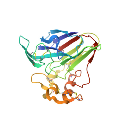
    Thaumatin, an intensely sweet-tasting protein, elicits a sweet taste sensation at 50 nM. Here the X-ray crystallographic structure of one of its variants, thaumatin II, was determined at a resolution of 1.27 Å. Overall structure of thaumatin II is similar to thaumatin I, but a slight shift of the Cα atom of G96 in thaumatin II was observed. Furthermore, the side chain of residue 67 in thaumatin II is highly disordered. Since residue 67 is one of two residues critical to the sweetness of thaumatin, the present results suggested that the critical positive charges at positions 67 and 82 are disordered and the flexibility and fluctuation of these side chains would be suitable for interaction of thaumatin molecules with sweet receptors.


  • Organizational Affiliation

    Division of Food Science and Biotechnology, Graduate School of Agriculture, Kyoto University, Uji, Kyoto 611-0011, Japan. t2masuda@kais.kyoto-u.ac.jp


Macromolecules
Find similar proteins by:  (by identity cutoff)  |  3D Structure
Entity ID: 1
MoleculeChains Sequence LengthOrganismDetailsImage
Thaumatin-2207Thaumatococcus danielliiMutation(s): 0 
UniProt
Find proteins for P02884 (Thaumatococcus daniellii)
Explore P02884 
Go to UniProtKB:  P02884
Entity Groups  
Sequence Clusters30% Identity50% Identity70% Identity90% Identity95% Identity100% Identity
UniProt GroupP02884
Sequence Annotations
Expand
  • Reference Sequence
Experimental Data & Validation

Experimental Data

  • Method: X-RAY DIFFRACTION
  • Resolution: 1.27 Å
  • R-Value Free: 0.148 
  • R-Value Observed: 0.112 
  • Space Group: P 41 21 2
Unit Cell:
Length ( Å )Angle ( ˚ )
a = 57.769α = 90
b = 57.769β = 90
c = 150.126γ = 90
Software Package:
Software NamePurpose
SHELXmodel building
SHELXL-97refinement
HKL-2000data reduction
HKL-2000data scaling
SHELXphasing

Structure Validation

View Full Validation Report


Entry History 

Deposition Data

Revision History  (Full details and data files)

  • Version 1.0: 2011-07-27
    Type: Initial release