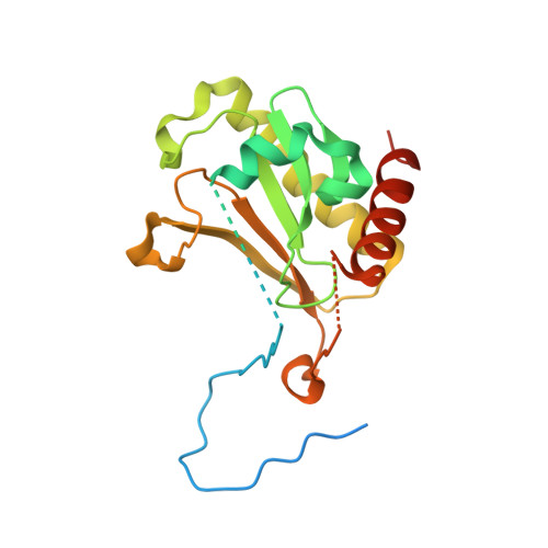
Structural insights into the novel diadenosine 5',5-P1,P4-tetraphosphate phosphorylase from Mycobacterium tuberculosis H37Rv
Mori, S., Shibayama, K., Wachino, J., Arakawa, Y.(2011) J Mol Biol 410: 93-104
- PubMed: 21565198
- DOI: https://doi.org/10.1016/j.jmb.2011.04.059
- Primary Citation of Related Structures:
3ANO - PubMed Abstract:
Rv2613c is a diadenosine 5',5‴-P(1),P(4)-tetraphosphate (Ap(4)A) phosphorylase from Mycobacterium tuberculosis H37Rv. Sequence analysis suggests that Rv2613c belongs to the histidine triad (HIT) motif superfamily, which includes HIT family diadenosine polyphosphate (Ap(n)A) hydrolases and Ap(4)A phosphorylases. However, the amino acid sequence of Rv2613c is more similar to that of HIT family Ap(n)A hydrolases than to that of typical Ap(4)A phosphorylases. Here, we report the crystal structure of Rv2613c, which is the first structure of a protein with Ap(n)A phosphorylase activity, and characterized the structural basis of its catalytic activity. Our results showed that the structure of Rv2613c is similar to those of other HIT superfamily proteins. However, Asn139, Gly146, and Ser147 in the active site of Rv2613c replace the corresponding Gln, Gln, and Thr residues that are normally found in HIT family Ap(n)A hydrolases. Furthermore, analyses of Rv2613c mutants revealed that Asn139, Gly146, and Ser147 are important active-site residues and that Asn139 has a critical role in catalysis. The position of Gly146 might influence the phosphorylase activity. In addition, the tetrameric structure of Rv2613c and the presence of Trp160 might be essential for the formation of the Ap(4)A binding site. These structural insights into Rv2613c may facilitate the development of novel structure-based inhibitors for treating tuberculosis.
Organizational Affiliation:
Department of Bacteriology II, National Institute of Infectious Diseases, 4-7-1 Gakuen, Musashi-Murayama-shi, Tokyo 208-0011, Japan. mshige@nig.go.jp