Crystal Structure of Clostridium perfringens Enterotoxin Displays Features of {beta}-Pore-forming Toxins
Kitadokoro, K., Nishimura, K., Kamitani, S., Fukui-Miyazaki, A., Toshima, H., Abe, H., Kamata, Y., Sugita-Konishi, Y., Yamamoto, S., Karatani, H., Horiguchi, Y.(2011) J Biol Chem 286: 19549-19555
- PubMed: 21489981
- DOI: https://doi.org/10.1074/jbc.M111.228478
- Primary Citation of Related Structures:
3AM2 - PubMed Abstract:
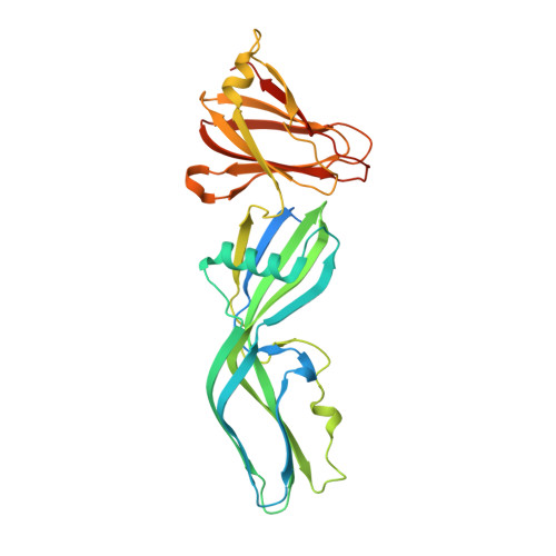
Clostridium perfringens enterotoxin (CPE) is a cause of food poisoning and is considered a pore-forming toxin, which damages target cells by disrupting the selective permeability of the plasma membrane. However, the pore-forming mechanism and the structural characteristics of the pores are not well documented. Here, we present the structure of CPE determined by x-ray crystallography at 2.0 Å. The overall structure of CPE displays an elongated shape, composed of three distinct domains, I, II, and III. Domain I corresponds to the region that was formerly referred to as C-CPE, which is responsible for binding to the specific receptor claudin. Domains II and III comprise a characteristic module, which resembles those of β-pore-forming toxins such as aerolysin, C. perfringens ε-toxin, and Laetiporus sulfureus hemolytic pore-forming lectin. The module is mainly made up of β-strands, two of which span its entire length. Domain II and domain III have three short β-strands each, by which they are distinguished. In addition, domain II has an α-helix lying on the β-strands. The sequence of amino acids composing the α-helix and preceding β-strand demonstrates an alternating pattern of hydrophobic residues that is characteristic of transmembrane domains forming β-barrel-made pores. These structural features imply that CPE is a β-pore-forming toxin. We also hypothesize that the transmembrane domain is inserted into the membrane upon the buckling of the two long β-strands spanning the module, a mechanism analogous to that of the cholesterol-dependent cytolysins.
Organizational Affiliation:
Graduate School of Science and Technology, Department of Biomolecular Engineering, Kyoto Institute of Technology, Sakyo-ku, Kyoto, Japan.