


Salt-induced conformational change of salt-tolerant glutaminase from Micrococcus luteus K-3
Yoshimune, K., Shirakihara, Y., Yumoto, I.To be published.
Experimental Data Snapshot
wwPDB Validation 3D Report Full Report

Entity ID: 1 | |||||
|---|---|---|---|---|---|
| Molecule | Chains | Sequence Length | Organism | Details | Image |
| Glutaminase 1 | 327 | Bacillus subtilis | Mutation(s): 0 Gene Names: Glutaminase EC: 3.5.1.2 | 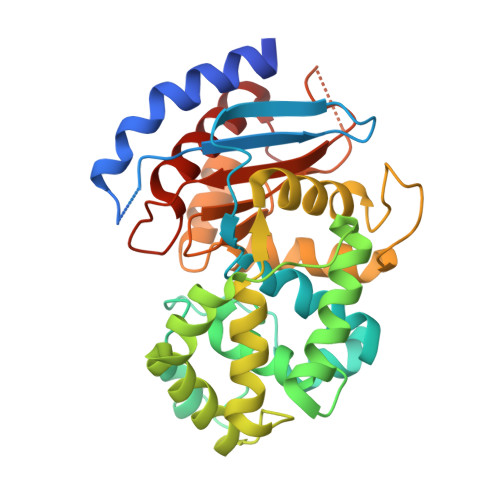 | |
UniProt | |||||
Find proteins for O31465 (Bacillus subtilis (strain 168)) Explore O31465 Go to UniProtKB: O31465 | |||||
Entity Groups | |||||
| Sequence Clusters | 30% Identity50% Identity70% Identity90% Identity95% Identity100% Identity | ||||
| UniProt Group | O31465 | ||||
Sequence AnnotationsExpand | |||||
| |||||
| Length ( Å ) | Angle ( ˚ ) |
|---|---|
| a = 70.837 | α = 90 |
| b = 179.965 | β = 90 |
| c = 51.878 | γ = 90 |
| Software Name | Purpose |
|---|---|
| ADSC | data collection |
| AMoRE | phasing |
| CNS | refinement |
| MOSFLM | data reduction |
| SCALA | data scaling |
RCSB PDB (citation) is hosted by
RCSB PDB is a member of the

