First crystal structure of L-lysine 6-dehydrogenase as an NAD-dependent amine dehydrogenase.
Yoneda, K., Fukuda, J., Sakuraba, H., Ohshima, T.(2010) J Biol Chem 285: 8444-8453
- PubMed: 20056607
- DOI: https://doi.org/10.1074/jbc.M109.084384
- Primary Citation of Related Structures:
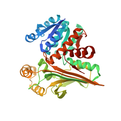
3ABI - PubMed Abstract:
A gene encoding an L-lysine dehydrogenase was identified in the hyperthermophilic archaeon Pyrococcus horikoshii. The gene was overexpressed in Escherichia coli, and its product was purified and characterized. The expressed enzyme is the most thermostable L-lysine dehydrogenase yet described, with a half-life of 180 min at 100 degrees C. The product of the enzyme's catalytic activity is Delta(1)-piperideine-6-carboxylate, which makes this enzyme an L-lysine 6-dehydrogenase (EC 1.4.1.18) that catalyzes the reductive deamination of the epsilon- amino group and a type of NAD-dependent amine dehydrogenase. The three-dimensional structure of the enzyme was determined using the mercury-based multiple-wavelength anomalous dispersion method at a resolution of 2.44 A in the presence of NAD and sulfate ion. The asymmetric unit consisted of two subunits, and a crystallographic 2-fold axis generated the functional dimer. Each monomer consisted of a Rossmann fold domain and a C-terminal catalytic domain, and the fold of the catalytic domain showed similarity to that of saccharopine reductase. Notably, the structures of subunits A and B differed significantly. In subunit A, the active site contained a sulfate ion that was not seen in subunit B. Consequently, subunit A adopted a closed conformation, whereas subunit B adopted an open one. In each subunit, one NAD molecule was bound to the active site in an anti-conformation, indicating that the enzyme makes use of pro-R-specific hydride transfer between the two hydrides at C-4 of NADH (type A specificity). This is the first description of the three-dimensional structure of l-lysine 6-dehydrogenase as an NAD-dependent amine dehydrogenase.
Organizational Affiliation:
Department of Bioscience, School of Agriculture, Tokai University, Aso, Kumamoto 869-1404, Japan.