3A9E

Crystal structure of a mixed agonist-bound RAR-alpha and antagonist-bound RXR-alpha heterodimer ligand binding domains


Experimental Data Snapshot

  • Method: X-RAY DIFFRACTION
  • Resolution: 2.75 Å
  • R-Value Free: 0.264 
  • R-Value Work: 0.201 
  • R-Value Observed: 0.204 

wwPDB Validation   3D Report Full Report


Ligand Structure Quality Assessment 


This is version 1.3 of the entry. See complete history


Literature

The Phantom Effect of the Rexinoid LG100754: structural and functional insights

Sato, Y.Ramalanjaona, N.Huet, T.Potier, N.Osz, J.Antony, P.Peluso-Iltis, C.Poussin-Courmontagne, P.Ennifar, E.Mely, Y.Dejaegere, A.Moras, D.Rochel, N.

(2010) PLoS One 5: e15119-e15119

  • DOI: https://doi.org/10.1371/journal.pone.0015119
  • Primary Citation of Related Structures:  
    3A9E

  • PubMed Abstract: 

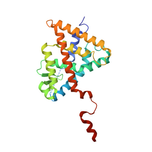
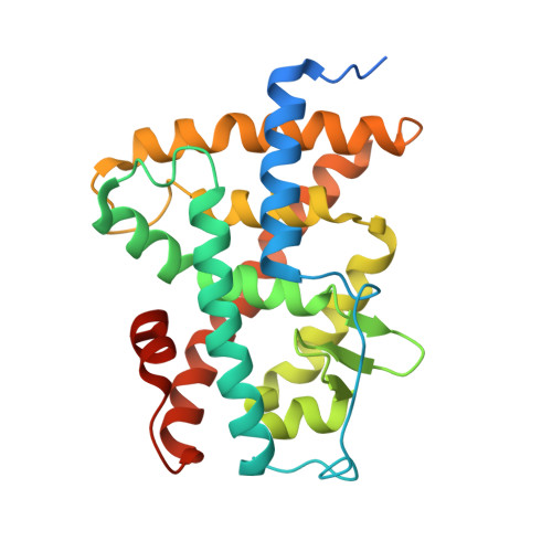
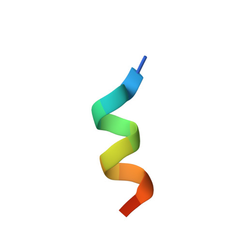
    Retinoic acid receptors (RARs) and Retinoid X nuclear receptors (RXRs) are ligand-dependent transcriptional modulators that execute their biological action through the generation of functional heterodimers. RXR acts as an obligate dimer partner in many signalling pathways, gene regulation by rexinoids depending on the liganded state of the specific heterodimeric partner. To address the question of the effect of rexinoid antagonists on RAR/RXR function, we solved the crystal structure of the heterodimer formed by the ligand binding domain (LBD) of the RARα bound to its natural agonist ligand (all-trans retinoic acid, atRA) and RXRα bound to a rexinoid antagonist (LG100754). We observed that RARα exhibits the canonical agonist conformation and RXRα an antagonist one with the C-terminal H12 flipping out to the solvent. Examination of the protein-LG100754 interactions reveals that its propoxy group sterically prevents the H12 associating with the LBD, without affecting the dimerization or the active conformation of RAR. Although LG100754 has been reported to act as a 'phantom ligand' activating RAR in a cellular context, our structural data and biochemical assays demonstrate that LG100754 mediates its effect as a full RXR antagonist. Finally we show that the 'phantom ligand effect' of the LG100754 is due to a direct binding of the ligand to RAR that stabilizes coactivator interactions thus accounting for the observed transcriptional activation of RAR/RXR.


  • Organizational Affiliation

    Département de Biologie et de Génomique Structurales, Institut de Génétique et de Biologie Moléculaire et Cellulaire (IGBMC), Institut National de Santé et de Recherche Médicale (INSERM) U964/Centre National de Recherche Scientifique (CNRS) UMR 1704/Université de Strasbourg, Illkirch, France.


Macromolecules
Find similar proteins by:  (by identity cutoff)  |  3D Structure
Entity ID: 1
MoleculeChains Sequence LengthOrganismDetailsImage
Retinoic acid receptor RXR-alpha240Mus musculusMutation(s): 0 
Gene Names: RxraNr2b1
UniProt
Find proteins for P28700 (Mus musculus)
Explore P28700 
Go to UniProtKB:  P28700
Entity Groups  
Sequence Clusters30% Identity50% Identity70% Identity90% Identity95% Identity100% Identity
UniProt GroupP28700
Sequence Annotations
Expand
  • Reference Sequence
Find similar proteins by:  (by identity cutoff)  |  3D Structure
Entity ID: 2
MoleculeChains Sequence LengthOrganismDetailsImage
Retinoic acid receptor alpha269Homo sapiensMutation(s): 0 
Gene Names: RARANR1B1
UniProt & NIH Common Fund Data Resources
Find proteins for P10276 (Homo sapiens)
Explore P10276 
Go to UniProtKB:  P10276
PHAROS:  P10276
GTEx:  ENSG00000131759 
Entity Groups  
Sequence Clusters30% Identity50% Identity70% Identity90% Identity95% Identity100% Identity
UniProt GroupP10276
Sequence Annotations
Expand
  • Reference Sequence

Find similar proteins by:  Sequence   |   3D Structure  

Entity ID: 3
MoleculeChains Sequence LengthOrganismDetailsImage
13-mer (LXXLL motif) from Nuclear receptor coactivator 2C [auth I]13Homo sapiensMutation(s): 0 
UniProt & NIH Common Fund Data Resources
Find proteins for Q15596 (Homo sapiens)
Explore Q15596 
Go to UniProtKB:  Q15596
PHAROS:  Q15596
GTEx:  ENSG00000140396 
Entity Groups  
Sequence Clusters30% Identity50% Identity70% Identity90% Identity95% Identity100% Identity
UniProt GroupQ15596
Sequence Annotations
Expand
  • Reference Sequence
Small Molecules
Ligands 2 Unique
IDChains Name / Formula / InChI Key2D Diagram3D Interactions
754
Query on 754

Download Ideal Coordinates CCD File 
D [auth A](2E,4E,6Z)-3-methyl-7-(5,5,8,8-tetramethyl-3-propoxy-5,6,7,8-tetrahydronaphthalen-2-yl)octa-2,4,6-trienoic acid
C26 H36 O3
HNODNXQAYXJFMQ-LQUSFLDPSA-N
REA
Query on REA

Download Ideal Coordinates CCD File 
E [auth B]RETINOIC ACID
C20 H28 O2
SHGAZHPCJJPHSC-YCNIQYBTSA-N
Binding Affinity Annotations 
IDSourceBinding Affinity
754 BindingDB:  3A9E Ki: min: 8, max: 2587 (nM) from 5 assay(s)
EC50: min: 2, max: 13 (nM) from 5 assay(s)
REA BindingDB:  3A9E IC50: min: 1000, max: 5.00e+4 (nM) from 5 assay(s)
Experimental Data & Validation

Experimental Data

  • Method: X-RAY DIFFRACTION
  • Resolution: 2.75 Å
  • R-Value Free: 0.264 
  • R-Value Work: 0.201 
  • R-Value Observed: 0.204 
  • Space Group: P 43 21 2
Unit Cell:
Length ( Å )Angle ( ˚ )
a = 105.3α = 90
b = 105.3β = 90
c = 111.338γ = 90
Software Package:
Software NamePurpose
MxCuBEdata collection
AMoREphasing
PHENIXrefinement
HKL-2000data reduction
HKL-2000data scaling

Structure Validation

View Full Validation Report


Ligand Structure Quality Assessment 


Entry History 

Deposition Data

Revision History  (Full details and data files)

  • Version 1.0: 2010-10-06
    Type: Initial release
  • Version 1.1: 2011-07-13
    Changes: Version format compliance
  • Version 1.2: 2013-10-30
    Changes: Database references
  • Version 1.3: 2023-11-01
    Changes: Data collection, Database references, Derived calculations, Refinement description