3A51

Structure of cytochrome P450 Vdh mutant (Vdh-K1) obtained by directed evolution with bound 25-hydroxyvitamin D3


Experimental Data Snapshot

  • Method: X-RAY DIFFRACTION
  • Resolution: 2.00 Å
  • R-Value Free: 0.234 
  • R-Value Work: 0.197 
  • R-Value Observed: 0.199 

wwPDB Validation   3D Report Full Report


Ligand Structure Quality Assessment 


This is version 1.2 of the entry. See complete history


Literature

Structural evidence for enhancement of sequential vitamin D3 hydroxylation activities by directed evolution of cytochrome P450 vitamin D3 hydroxylase

Yasutake, Y.Fujii, Y.Nishioka, T.Cheon, W.K.Arisawa, A.Tamura, T.

(2010) J Biol Chem 285: 31193-31201

  • DOI: https://doi.org/10.1074/jbc.M110.147009
  • Primary Citation of Related Structures:  
    3A4G, 3A4H, 3A4Z, 3A50, 3A51

  • PubMed Abstract: 

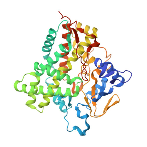
    Vitamin D(3) hydroxylase (Vdh) isolated from actinomycete Pseudonocardia autotrophica is a cytochrome P450 (CYP) responsible for the biocatalytic conversion of vitamin D(3) (VD(3)) to 1α,25-dihydroxyvitamin D(3) (1α,25(OH)(2)VD(3)) by P. autotrophica. Although its biological function is unclear, Vdh is capable of catalyzing the two-step hydroxylation of VD(3), i.e. the conversion of VD(3) to 25-hydroxyvitamin D(3) (25(OH)VD(3)) and then of 25(OH)VD(3) to 1α,25(OH)(2)VD(3), a hormonal form of VD(3). Here we describe the crystal structures of wild-type Vdh (Vdh-WT) in the substrate-free form and of the highly active quadruple mutant (Vdh-K1) generated by directed evolution in the substrate-free, VD(3)-bound, and 25(OH)VD(3)-bound forms. Vdh-WT exhibits an open conformation with the distal heme pocket exposed to the solvent both in the presence and absence of a substrate, whereas Vdh-K1 exhibits a closed conformation in both the substrate-free and substrate-bound forms. The results suggest that the conformational equilibrium was largely shifted toward the closed conformation by four amino acid substitutions scattered throughout the molecule. The substrate-bound structure of Vdh-K1 accommodates both VD(3) and 25(OH)VD(3) but in an anti-parallel orientation. The occurrence of the two secosteroid binding modes accounts for the regioselective sequential VD(3) hydroxylation activities. Moreover, these structures determined before and after directed evolution, together with biochemical and spectroscopic data, provide insights into how directed evolution has worked for significant enhancement of both the VD(3) 25-hydroxylase and 25(OH)VD(3) 1α-hydroxylase activities.


  • Organizational Affiliation

    Bioproduction Research Institute, National Institute of Advanced Industrial Science and Technology, Sapporo 062-8517, Japan.


Macromolecules
Find similar proteins by:  (by identity cutoff)  |  3D Structure
Entity ID: 1
MoleculeChains Sequence LengthOrganismDetailsImage
Vitamin D hydroxylase
A, B, C, D, E
411Pseudonocardia autotrophicaMutation(s): 4 
Gene Names: vdh
UniProt
Find proteins for C4B644 (Pseudonocardia autotrophica)
Explore C4B644 
Go to UniProtKB:  C4B644
Entity Groups  
Sequence Clusters30% Identity50% Identity70% Identity90% Identity95% Identity100% Identity
UniProt GroupC4B644
Sequence Annotations
Expand
  • Reference Sequence
Small Molecules
Ligands 5 Unique
IDChains Name / Formula / InChI Key2D Diagram3D Interactions
HEM
Query on HEM

Download Ideal Coordinates CCD File 
F [auth A],
N [auth B],
Q [auth C],
V [auth D],
Z [auth E]
PROTOPORPHYRIN IX CONTAINING FE
C34 H32 Fe N4 O4
KABFMIBPWCXCRK-RGGAHWMASA-L
VDY
Query on VDY

Download Ideal Coordinates CCD File 
AA [auth E],
G [auth A],
O [auth B],
R [auth C],
W [auth D]
3-{2-[1-(5-HYDROXY-1,5-DIMETHYL-HEXYL)-7A-METHYL-OCTAHYDRO-INDEN-4-YLIDENE]-ETHYLIDENE}-4-METHYLENE-CYCLOHEXANOL
C27 H44 O2
JWUBBDSIWDLEOM-DTOXIADCSA-N
GOL
Query on GOL

Download Ideal Coordinates CCD File 
K [auth A],
M [auth A]
GLYCEROL
C3 H8 O3
PEDCQBHIVMGVHV-UHFFFAOYSA-N
ACT
Query on ACT

Download Ideal Coordinates CCD File 
CA [auth E]
DA [auth E]
L [auth A]
P [auth B]
T [auth C]
CA [auth E],
DA [auth E],
L [auth A],
P [auth B],
T [auth C],
U [auth C],
Y [auth D]
ACETATE ION
C2 H3 O2
QTBSBXVTEAMEQO-UHFFFAOYSA-M
CA
Query on CA

Download Ideal Coordinates CCD File 
BA [auth E]
H [auth A]
I [auth A]
J [auth A]
S [auth C]
BA [auth E],
H [auth A],
I [auth A],
J [auth A],
S [auth C],
X [auth D]
CALCIUM ION
Ca
BHPQYMZQTOCNFJ-UHFFFAOYSA-N
Experimental Data & Validation

Experimental Data

  • Method: X-RAY DIFFRACTION
  • Resolution: 2.00 Å
  • R-Value Free: 0.234 
  • R-Value Work: 0.197 
  • R-Value Observed: 0.199 
  • Space Group: P 21 21 21
Unit Cell:
Length ( Å )Angle ( ˚ )
a = 77.173α = 90
b = 171.8β = 90
c = 189.143γ = 90
Software Package:
Software NamePurpose
ADSCdata collection
MOLREPphasing
REFMACrefinement
HKL-2000data reduction
HKL-2000data scaling

Structure Validation

View Full Validation Report


Ligand Structure Quality Assessment 


Entry History 

Deposition Data

Revision History  (Full details and data files)

  • Version 1.0: 2010-07-28
    Type: Initial release
  • Version 1.1: 2011-07-13
    Changes: Version format compliance
  • Version 1.2: 2023-11-01
    Changes: Data collection, Database references, Derived calculations, Refinement description