3A1Q

Crystal structure of the mouse RAP80 UIMs in complex with Lys63-linked di-ubiquitin


Experimental Data Snapshot

  • Method: X-RAY DIFFRACTION
  • Resolution: 2.20 Å
  • R-Value Free: 0.268 
  • R-Value Work: 0.216 
  • R-Value Observed: 0.216 

wwPDB Validation   3D Report Full Report


This is version 1.3 of the entry. See complete history


Literature

Structural basis for specific recognition of Lys 63-linked polyubiquitin chains by tandem UIMs of RAP80

Sato, Y.Yoshikawa, A.Mimura, H.Yamashita, M.Yamagata, A.Fukai, S.

(2009) EMBO J 28: 2461-2468

  • DOI: https://doi.org/10.1038/emboj.2009.160
  • Primary Citation of Related Structures:  
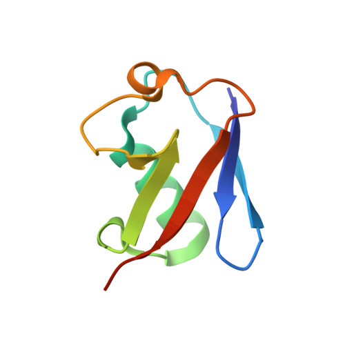
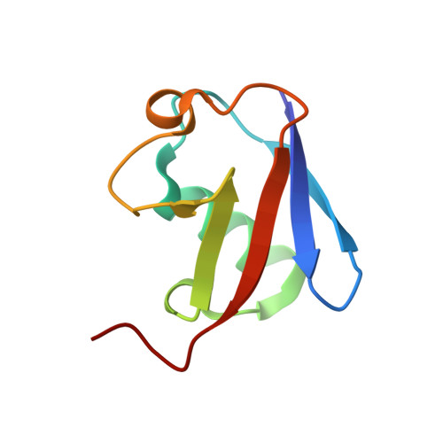
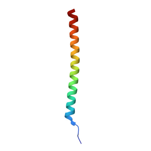
    3A1Q

  • PubMed Abstract: 

    RAP80 has a key role in the recruitment of the Abraxas-BRCC36-BRCA1-BARD1 complex to DNA-damage foci for DNA repair through specific recognition of Lys 63-linked polyubiquitinated proteins by its tandem ubiquitin-interacting motifs (UIMs). Here, we report the crystal structure of the RAP80 tandem UIMs (RAP80-UIM1-UIM2) in complex with Lys 63-linked di-ubiquitin at 2.2 A resolution. The two UIMs, UIM1 and UIM2, and the alpha-helical inter-UIM region together form a continuous 60 A-long alpha-helix. UIM1 and UIM2 bind to the proximal and distal ubiquitin moieties, respectively. Both UIM1 and UIM2 of RAP80 recognize an Ile 44-centered hydrophobic patch on ubiquitin but neither UIM interacts with the Lys 63-linked isopeptide bond. Our structure suggests that the inter-UIM region forms a 12 A-long alpha-helix that ensures that the UIMs are arranged to enable specific binding of Lys 63-linked di-ubiquitin. This was confirmed by pull-down analyses using RAP80-UIM1-UIM2 mutants of various length inter-UIM regions. Further, we show that the Epsin1 tandem UIM, which has an inter-UIM region similar to that of RAP80-UIM1-UIM2, also selectively binds Lys 63-linked di-ubiquitin.


  • Organizational Affiliation

    Structural Biology Laboratory, Life Science Division, Synchrotron Radiation Research Organization and Institute of Molecular and Cellular Biosciences, The University of Tokyo, Tokyo, Japan.


Macromolecules
Find similar proteins by:  (by identity cutoff)  |  3D Structure
Entity ID: 1
MoleculeChains Sequence LengthOrganismDetailsImage
Ubiquitin
A, D
77Mus musculusMutation(s): 1 
Gene Names: Rps27aUba80Ubcep1
UniProt & NIH Common Fund Data Resources
Find proteins for P0CG50 (Mus musculus)
Explore P0CG50 
Go to UniProtKB:  P0CG50
IMPC:  MGI:98889
Entity Groups  
Sequence Clusters30% Identity50% Identity70% Identity90% Identity95% Identity100% Identity
UniProt GroupP0CG50
Sequence Annotations
Expand
  • Reference Sequence
Find similar proteins by:  (by identity cutoff)  |  3D Structure
Entity ID: 2
MoleculeChains Sequence LengthOrganismDetailsImage
Ubiquitin
B, E
76Mus musculusMutation(s): 1 
Gene Names: Rps27aUba80Ubcep1
UniProt & NIH Common Fund Data Resources
Find proteins for P0CG50 (Mus musculus)
Explore P0CG50 
Go to UniProtKB:  P0CG50
IMPC:  MGI:98889
Entity Groups  
Sequence Clusters30% Identity50% Identity70% Identity90% Identity95% Identity100% Identity
UniProt GroupP0CG50
Sequence Annotations
Expand
  • Reference Sequence
Find similar proteins by:  (by identity cutoff)  |  3D Structure
Entity ID: 3
MoleculeChains Sequence LengthOrganismDetailsImage
Ubiquitin interaction motif-containing protein 1
C, F
45Mus musculusMutation(s): 0 
Gene Names: Uimc1Rip110Rxrip110
UniProt
Find proteins for Q5U5Q9 (Mus musculus)
Explore Q5U5Q9 
Go to UniProtKB:  Q5U5Q9
Entity Groups  
Sequence Clusters30% Identity50% Identity70% Identity90% Identity95% Identity100% Identity
UniProt GroupQ5U5Q9
Sequence Annotations
Expand
  • Reference Sequence
Experimental Data & Validation

Experimental Data

  • Method: X-RAY DIFFRACTION
  • Resolution: 2.20 Å
  • R-Value Free: 0.268 
  • R-Value Work: 0.216 
  • R-Value Observed: 0.216 
  • Space Group: P 21 21 21
Unit Cell:
Length ( Å )Angle ( ˚ )
a = 65.235α = 90
b = 74.81β = 90
c = 83.031γ = 90
Software Package:
Software NamePurpose
CNSrefinement
ADSCdata collection
HKL-2000data reduction
HKL-2000data scaling
MOLREPphasing

Structure Validation

View Full Validation Report


Entry History 

Deposition Data

Revision History  (Full details and data files)

  • Version 1.0: 2009-07-21
    Type: Initial release
  • Version 1.1: 2011-07-13
    Changes: Version format compliance
  • Version 1.2: 2021-11-10
    Changes: Database references
  • Version 1.3: 2023-11-01
    Changes: Data collection, Refinement description