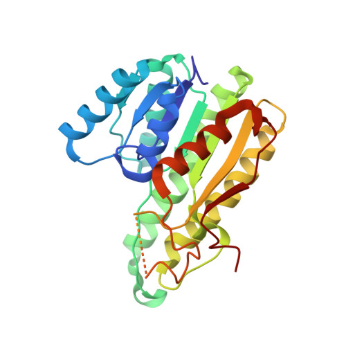
Closed complex of the D-3-hydroxybutyrate dehydrogenase induced by an enantiomeric competitive inhibitor.
Nakashima, K., Ito, K., Nakajima, Y., Yamazawa, R., Miyakawa, S., Yoshimoto, T.(2009) J Biochem 145: 467-479
- PubMed: 19122202
- DOI: https://doi.org/10.1093/jb/mvn186
- Primary Citation of Related Structures:
2ZTL, 2ZTM, 2ZTU, 2ZTV - PubMed Abstract:
D-3-Hydroxybutyrate dehydrogenase (HBDH) from Pseudomonas fragi showed a strict stereospecificity to the d-enantiomer of 3-hydroxybutyrate (d-3-HB) as a substrate. The l-enantiomer acts as a competitive inhibitor, with a K(i) value comparable to the K(m) value for d-3-HB. We have determined the crystal structures of the ternary complex of HBDH-NAD(+)-l-3-HB and the binary complex of HBDH-NAD(+). The former structure showed a so-called closed-form conformation, which is considered an active form for catalysis, while the latter stayed mostly in a open-form conformation. The determined structures along with the site-directed mutagenesis confirmed the substrate recognition mechanism that we proposed previously. The hydrogen bonding interaction between Gln196, located in the moving helix, and the carboxyl group of the substrate/inhibitor is important for the stable ternary complex formation. Finally, the crystal structures of the Thr190 mutants, T190S and T190A, indicate that the Thr190 is a key residue for the open-closed conformational change. T190S retained 37% of the activity. In T190A, however, the activity decreased to 0.1% that of the wild-type enzyme. Fixing the position of the hydroxyl group of Thr190 to form hydrogen bonds to the pyrophosphate moiety and the carboxamide of NAD(+) seems to be a significant factor for the open-closed conformational change.
Organizational Affiliation:
Department of Molecular Medicinal Sciences, Graduate School of Biomedical Sciences, Nagasaki University, 1-14 Bunkyo-machi, Nagasaki 852-8521, Japan.


