Crystal structure of the parasporin-2 Bacillus thuringiensis toxin that recognizes cancer cells
Akiba, T., Abe, Y., Kitada, S., Kusaka, Y., Ito, A., Ichimatsu, T., Katayama, H., Akao, T., Higuchi, K., Mizuki, E., Ohba, M., Kanai, R., Harata, K.(2009) J Mol Biol 386: 121-133
- PubMed: 19094993
- DOI: https://doi.org/10.1016/j.jmb.2008.12.002
- Primary Citation of Related Structures:
2ZTB - PubMed Abstract:
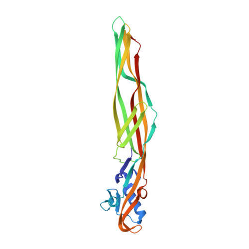
Parasporin-2 is a protein toxin that is isolated from parasporal inclusions of the Gram-positive bacterium Bacillus thuringiensis. Although B. thuringiensis is generally known as a valuable source of insecticidal toxins, parasporin-2 is not insecticidal, but has a strong cytocidal activity in liver and colon cancer cells. The 37-kDa inactive nascent protein is proteolytically cleaved to the 30-kDa active form that loses both the N-terminal and the C-terminal segments. Accumulated cytological and biochemical observations on parasporin-2 imply that the protein is a pore-forming toxin. To confirm the hypothesis, we have determined the crystal structure of its active form at a resolution of 2.38 A. The protein is unusually elongated and mainly comprises long beta-strands aligned with its long axis. It is similar to aerolysin-type beta-pore-forming toxins, which strongly reinforce the pore-forming hypothesis. The molecule can be divided into three domains. Domain 1, comprising a small beta-sheet sandwiched by short alpha-helices, is probably the target-binding module. Two other domains are both beta-sandwiches and thought to be involved in oligomerization and pore formation. Domain 2 has a putative channel-forming beta-hairpin characteristic of aerolysin-type toxins. The surface of the protein has an extensive track of exposed side chains of serine and threonine residues. The track might orient the molecule on the cell membrane when domain 1 binds to the target until oligomerization and pore formation are initiated. The beta-hairpin has such a tight structure that it seems unlikely to reform as postulated in a recent model of pore formation developed for aerolysin-type toxins. A safety lock model is proposed as an inactivation mechanism by the N-terminal inhibitory segment.
Organizational Affiliation:
Biological Information Research Center, AIST, Tsukuba, Ibaraki 305-8566, Japan. toshi-akiba@aist.go.jp