2ZJ9

X-ray crystal structure of AmpC beta-Lactamase (AmpC(D)) from an Escherichia coli with a Tripeptide Deletion (Gly286 Ser287 Asp288) on the H10 Helix


Experimental Data Snapshot

  • Method: X-RAY DIFFRACTION
  • Resolution: 1.70 Å
  • R-Value Free: 0.206 
  • R-Value Work: 0.163 
  • R-Value Observed: 0.165 

wwPDB Validation   3D Report Full Report


This is version 1.2 of the entry. See complete history


Literature

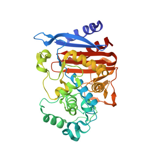
Structure of AmpC beta-lactamase (AmpCD) from an Escherichia coli clinical isolate with a tripeptide deletion (Gly286-Ser287-Asp288) in the H10 helix

Yamaguchi, Y.Sato, G.Yamagata, Y.Doi, Y.Wachino, J.Arakawa, Y.Matsuda, K.Kurosaki, H.

(2009) Acta Crystallogr Sect F Struct Biol Cryst Commun 65: 540-543

  • DOI: https://doi.org/10.1107/S1744309109014249
  • Primary Citation of Related Structures:  
    2ZJ9

  • PubMed Abstract: 

    The X-ray crystal structure of AmpC beta-lactamase (AmpC(D)) with a tripeptide deletion (Gly286-Ser287-Asp288) produced by Escherichia coli HKY28, a ceftazidime-resistant strain, was determined at a resolution of 1.7 A. The structure of AmpC(D) suggests that the tripeptide deletion at positions 286-288 located in the H10 helix causes a structural change of the Asn289-Asn294 region from the alpha-helix present in the native AmpC beta-lactamase of E. coli to a loop structure, which results in a widening of the substrate-binding site.


  • Organizational Affiliation

    Environmental Safety Center, Kumamoto University, Kumamoto, Japan. yyamagu@gpo.kumamoto-u.ac.jp


Macromolecules
Find similar proteins by:  (by identity cutoff)  |  3D Structure
Entity ID: 1
MoleculeChains Sequence LengthOrganismDetailsImage
AmpC
A, B
355Escherichia coliMutation(s): 0 
EC: 3.5.2.6
UniProt
Find proteins for Q76DI4 (Escherichia coli)
Explore Q76DI4 
Go to UniProtKB:  Q76DI4
Entity Groups  
Sequence Clusters30% Identity50% Identity70% Identity90% Identity95% Identity100% Identity
UniProt GroupQ76DI4
Sequence Annotations
Expand
  • Reference Sequence
Experimental Data & Validation

Experimental Data

  • Method: X-RAY DIFFRACTION
  • Resolution: 1.70 Å
  • R-Value Free: 0.206 
  • R-Value Work: 0.163 
  • R-Value Observed: 0.165 
  • Space Group: P 1
Unit Cell:
Length ( Å )Angle ( ˚ )
a = 47.065α = 82.62
b = 47.381β = 80.91
c = 81.461γ = 65.4
Software Package:
Software NamePurpose
REFMACrefinement
HKL-2000data collection
HKL-2000data reduction
HKL-2000data scaling
AMoREphasing

Structure Validation

View Full Validation Report


Entry History 

Deposition Data

Revision History  (Full details and data files)

  • Version 1.0: 2009-03-10
    Type: Initial release
  • Version 1.1: 2011-07-13
    Changes: Version format compliance
  • Version 1.2: 2023-11-01
    Changes: Data collection, Database references, Derived calculations, Refinement description