Substrate recognition mechanism of alpha-1,6-glucosidic linkage hydrolyzing enzyme, dextran glucosidase from Streptococcus mutans.
Hondoh, H., Saburi, W., Mori, H., Okuyama, M., Nakada, T., Matsuura, Y., Kimura, A.(2008) J Mol Biol 378: 911-920
- PubMed: 18395742
- DOI: https://doi.org/10.1016/j.jmb.2008.03.016
- Primary Citation of Related Structures:
2ZIC, 2ZID - PubMed Abstract:
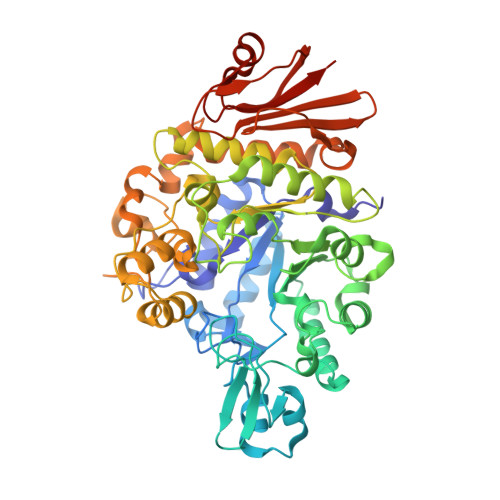
We have determined the crystal structure of Streptococcus mutans dextran glucosidase, which hydrolyzes the alpha-1,6-glucosidic linkage of isomaltooligosaccharides from their non-reducing ends to produce alpha-glucose. By using the mutant of catalytic acid Glu236-->Gln, its complex structure with the isomaltotriose, a natural substrate of this enzyme, has been determined. The enzyme has 536 amino acid residues and a molecular mass of 62,001 Da. The native and the complex structures were determined by the molecular replacement method and refined to 2.2 A resolution, resulting in a final R-factor of 18.3% for significant reflections in the native structure and 18.4% in the complex structure. The enzyme is composed of three domains, A, B and C, and has a (beta/alpha)(8)-barrel in domain A, which is common to the alpha-amylase family enzymes. Three catalytic residues are located at the bottom of the active site pocket and the bound isomaltotriose occupies subsites -1 to +2. The environment of the glucose residue at subsite -1 is similar to the environment of this residue in the alpha-amylase family. Hydrogen bonds between Asp60 and Arg398 and O4 atom of the glucose unit at subsite -1 accomplish recognition of the non-reducing end of the bound substrate. The side-chain atoms of Glu371 and Lys275 form hydrogen bonds with the O2 and O3 atoms of the glucose residue at subsite +1. The positions of atoms that compose the scissile alpha-1,6-glucosidic linkage (C1, O6 and C6 atoms) are identical with the positions of the atoms in the scissile alpha-1,4 linkage (C1, O4 and C4 atoms) of maltopentaose in the alpha-amylase structure from Bacillus subtilis. The comparison with the alpha-amylase suggests that Val195 of the dextran glucosidase and the corresponding residues of alpha-1,6-hydrolyzing enzymes participate in the determination of the substrate specificity of these enzymes.
Organizational Affiliation:
Research Faculty of Agriculture, Hokkaido University, Sapporo 060-8589, Japan. hondoh@abs.agr.hokudai.ac.jp