Crystal structure and molecular dynamics simulation of ubiquitin-like domain of murine parkin
Tomoo, K., Mukai, Y., In, Y., Miyagawa, H., Kitamura, K., Yamano, A., Shindo, H., Ishida, T.(2008) Biochim Biophys Acta 1784: 1059-1067
- PubMed: 18485927
- DOI: https://doi.org/10.1016/j.bbapap.2008.04.009
- Primary Citation of Related Structures:
2ZEQ - PubMed Abstract:
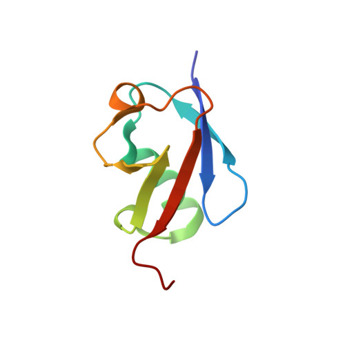
Parkin is the gene product identified as the major cause of autosomal recessive juvenile Parkinsonism (AR-JP). Parkin, a ubiquitin ligase E3, contains a unique ubiquitin-like domain in its N-terminus designated Uld which is assumed to be a interaction domain with the Rpn 10 subunit of 26S proteasome. To elucidate the structural and functional role of Uld in parkin at the atomic level, the X-ray crystal structure of murine Uld was determined and a molecular dynamics simulation of wild Uld and its five mutants (K27N, R33Q, R42P, K48A and V56E) identified from AR-JP patients was performed. Murine Uld consists of two alpha helices [Ile23-Arg33 (alpha1) and Val56-Gln57 (alpha2)] and five beta strands [Met1-Phe7 (beta1), Tyr11-Asp18 (beta2), Leu41-Phe45 (beta3), Lys48-Pro51 (beta4) and Ser65-Arg72 (beta5)] and its overall structure is essentially the same as that of human ubiquitin with a 1.22 A rmsd for the backbone atoms of residues 1-76; however, the sequential identity and similarity between both molecules are 32% and 63%, respectively. This close resemblance is due to the core structure built by same hydrogen bond formations between and within the backbone chains of alpha1 and beta1/2/5 secondary structure elements and by nearly the same hydrophobic interactions formed between the nonpolar amino acids of their secondary structures. The side chain NetaH of Lys27 on the alpha1 helix was crucial to the stabilization of the spatial orientations of beta3 and beta4 strands, possible binding region with Rpn 10 subunit, through three hydrogen bonds. The MD simulations showed the K27N and R33Q mutations increase the structural fluctuation of these beta strands including the alpha1 helix. Reversely, the V56E mutant restricted the spatial flexibility at the periphery of the short alpha2 helix by the interactions between the polar atoms of Glu56 and Ser19 residues. However, a large fluctuation of beta4 strand with respect to beta5 strand was induced in the R42P mutant, because of the impossibility of forming paired hydrogen bonds of Pro for Arg42 in wild Uld. The X-ray structure showed that the side chains of Asp39, Gln40 and Arg42 at the N-terminal periphery of beta3 strand protrude from the molecular surface of Uld and participate in hydrogen bonds with the polar residues of neighboring Ulds. Thus, the MD simulation suggests that the mutation substitution of Pro for Arg42 not only causes the large fluctuation of beta3 strand in the Uld but also leads to the loss of the ability of Uld to trap the Rpn 10 subunit. In contrast, the MD simulation of K48A mutant showed little influence on the beta3-beta4 loop structure, but a large fluctuation of Lys48 side chain, suggesting the importance of flexibility of this side chain for the interaction with the Rpn 10 subunit. The present results would be important in elucidating the impaired proteasomal binding mechanism of parkin in AR-JP.
Organizational Affiliation:
Department of Physical Chemistry, Osaka University of Pharmaceutical Sciences, Takatsuki, Osaka 569-1094, Japan. tomoo@gly.oups.ac.jp