Crystal structure of cytochrome P450 MoxA from Nonomuraea recticatena (CYP105)
Yasutake, Y., Imoto, N., Fujii, Y., Fujii, T., Arisawa, A., Tamura, T.(2007) Biochem Biophys Res Commun 361: 876-882
- PubMed: 17679139
- DOI: https://doi.org/10.1016/j.bbrc.2007.07.062
- Primary Citation of Related Structures:
2Z36 - PubMed Abstract:
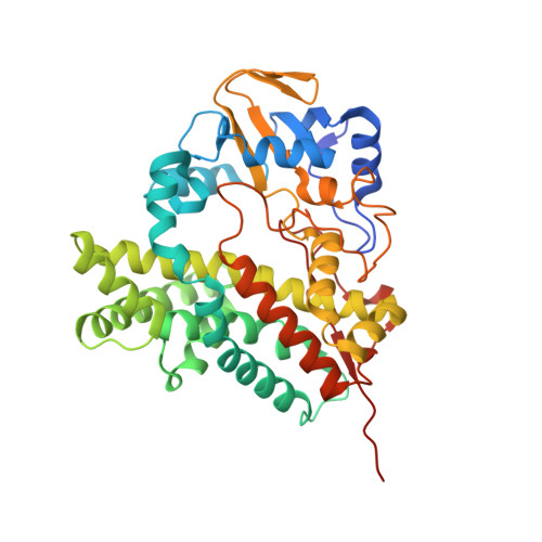
Cytochrome P450 MoxA (P450moxA) from a rare actinomycete Nonomuraea recticatena belongs to the CYP105 family and exhibits remarkably broad substrate specificity. Here, we demonstrate that P450moxA acts on several luciferin derivatives, which were originally identified as substrates of the human microsomal P450s. We also describe the crystal structure of P450moxA in substrate-free form. Structural comparison with various bacterial and human microsomal P450s reveals that the P450moxA structure is most closely related to that of the fungal nitric oxide reductase P450nor (CYP55A1). Final refined model of P450moxA comprises almost all the residues, including the "BC-loop" and "FG-loop" regions pivotal for substrate recognition, and the current structure thus defines a well-ordered substrate-binding pocket. Clear electron density map reveals that the MES molecule is bound to the substrate-binding site, and the sixth coordination position of the heme iron is not occupied by a water molecule, probably due to the presence of MES molecule in the vicinity of the heme. The unexpected binding of the MES molecule might reflect the ability of P450moxA to accommodate a broad range of structurally diverse compounds.
Organizational Affiliation:
Research Institute of Genome-based Biofactory, National Institute of Advanced Industrial Science and Technology, 2-17-2-1 Tsukisamu-Higashi, Toyohira-ku, Sapporo 062-8517, Japan.