


Crystal Structure of GDP-D-Mannose Dehydratase from Aquifex aeolicus VF5
Niwa, H., Kuramitsu, S., Yokoyama, S.To be published.
Experimental Data Snapshot
Entity ID: 1 | |||||
|---|---|---|---|---|---|
| Molecule | Chains | Sequence Length | Organism | Details | Image |
| GDP-D-mannose dehydratase | 345 | Aquifex aeolicus VF5 | Mutation(s): 0 Gene Names: rfbD EC: 4.2.1.47 | 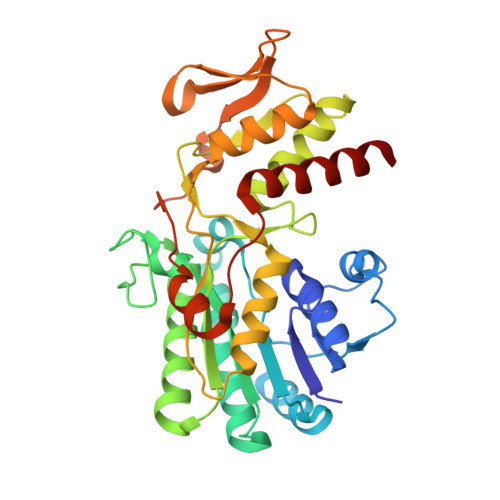 | |
UniProt | |||||
Find proteins for O67175 (Aquifex aeolicus (strain VF5)) Explore O67175 Go to UniProtKB: O67175 | |||||
Entity Groups | |||||
| Sequence Clusters | 30% Identity50% Identity70% Identity90% Identity95% Identity100% Identity | ||||
| UniProt Group | O67175 | ||||
Sequence AnnotationsExpand | |||||
| |||||
| Ligands 2 Unique | |||||
|---|---|---|---|---|---|
| ID | Chains | Name / Formula / InChI Key | 2D Diagram | 3D Interactions | |
| NDP Query on NDP | E [auth A], G [auth D] | NADPH DIHYDRO-NICOTINAMIDE-ADENINE-DINUCLEOTIDE PHOSPHATE C21 H30 N7 O17 P3 ACFIXJIJDZMPPO-NNYOXOHSSA-N |  | ||
| GDP Query on GDP | F [auth A], H [auth D] | GUANOSINE-5'-DIPHOSPHATE C10 H15 N5 O11 P2 QGWNDRXFNXRZMB-UUOKFMHZSA-N |  | ||
| Length ( Å ) | Angle ( ˚ ) |
|---|---|
| a = 58.799 | α = 90 |
| b = 151.164 | β = 108.6 |
| c = 85.934 | γ = 90 |
| Software Name | Purpose |
|---|---|
| REFMAC | refinement |
| HKL-2000 | data collection |
| HKL-2000 | data reduction |
| HKL-2000 | data scaling |
| CNS | phasing |
RCSB PDB (citation) is hosted by
RCSB PDB is a member of the

